Bioarchaeology International
Volume 7, Number 1: 1–31
DOI: 10.5744/bi.2022.0011
Received 22 February 2022
Revised 27 June 2022
Accepted 27 June 2022
Structural Violence and Physical Death at Tlatelolco: Selecting the Chronically Malnourished for Sacrifice at a Late Postclassic Mesoamerican City (1300–1521 CE)
Kelly E. Blevins,a,b* Madeline McGrane,b Josefina Mansilla Lory,c Salvador Guilliem Arroyo,d and Jane E. Buikstrab
ABSTRACT Human sacrifice in Mesoamerican cities was diverse and highly ritualized, and it remains incompletely understood. Knowing who was selected for ritual violence is essential for interpreting specialized mortuary deposits and furthering research on Mexica society. To understand the structure and variability of sacrificial and mortuary practices, we examine here three burial contexts from Tlatelolco, a densely populated city in the heart of the Triple Alliance. The interment contexts of Grupo Norte (n = 52) and Paso a Desnivel (n = 45) had been excavated from within the ceremonial center near the Tlatelolco Templo Mayor, and Atenantitech (n = 40) from a bordering calpulli or neighborhood. To establish which contexts are likely sacrificial deposits, we compare the age-at-death distributions, biological sex, and perimortem ritual trauma across these sites. We seek to understand if social status determined sacrificial inclusion by using metabolic and infectious disease as proxies for resource inequality. We find that the residential deposit approximates an attritional mortality distribution and that ceremonial center deposits primarily comprised non-adults, who also presented with significantly higher rates of metabolic and infectious disease than the non-adults from the residential site. Informed by previous studies and the ethnohistorical literature, we propose that impoverished individuals living on the margins of Mexica society were chosen as sacrificial victims. High prevalence of metabolic and infectious disease comorbidity indicates that these individuals endured long-term nutritional deficiency, apparently vitamin C. Further, variation in age, pathology, and perimortem treatment among ceremonial center deposits reveals the striking diversity of ritualized killings in a prominent Mexica city.
Keywords: human sacrifice; structural violence; Tlatelolco; Mesoamerica; Mexica; scurvy
El sacrificio en mesoamérica fue en extremo diverso, con muy diferentes y complejos rituales, saber quién y porqué fue seleccionado, para ser consagrados en un lugar específico es fundamental para interpretar los depósitos mortuorios y avanzar en la investigación sobre la sociedad mexica. Para comprender la estructura y variabilidad de las prácticas mortuorias, examinamos tres contextos de entierros de Tlatelolco, una ciudad densamente poblada localizada en el corazón de la Triple Alianza. Los contextos de entierro de Grupo Norte (n = 52) y Paso a Desnivel (n = 45) habían sido excavados dentro del centro ceremonial cerca del Templo Mayor de Tlatelolco, y Atenantitech (n = 40) de un barrio o calpulli limítrofe. Para confirmar y explicar qué contextos son depósitos de sacrificio, comparamos las distribuciones de edad biológica al morir, el sexo y el trauma ritual perimortem en estos restos humanos. Buscamos esclarecer si el estatus social determina la inclusión sacrificatoria mediante el análisis de enfermedades metabólicas e infecciosas como parámetros de una desigualdad de recursos. Encontramos que los individuos del sitio de enterramientos residencial se aproxima a una distribución de mortalidad por deterioro físico y que los depósitos de restos humanos del centro ceremonial estaban compuestos principalmente por sujetos no adultos que a su vez presentan tasas significativamente más altas de enfermedades metabólicas e infecciosas que los no adultos del sitio residencial. Con base en estudios previos y la literatura etnohistórica, proponemos que algunos individuos con una calidad de vida menoscabada que vivían en los márgenes de la sociedad mexica, fueron elegidos como víctimas de sacrificio. La alta prevalencia de comorbilidad de enfermedades metabólicas e infecciosas indica que estas personas soportaron durante un largo plazo deficiencias nutricionales, aparentemente de vitamina C. Además, la variación entre la edad, la patología y el tratamiento perimortem entre los individuos de los depósitos mortuorios del centro ceremonial revela una sorprendente diversidad de rituales sacrificiales en una ciudad mexica prominente.
Palabras claves: sacrificio humano; violencia estructural; Tlatelolco; Mesoamerica; Mexica; escorbuto
Mexica sacrificial rites were complex, distinctive, and predictable; human sacrifice was methodically practiced throughout an annual cycle of ceremonies and festivals. Human remains were offered to the gods as intricate ofrendas from contexts as grand as templos mayores to commonplace residential courtyards, bodies were butchered for ceremonial consumption, and skulls were organized into racks and columns called tzompantli as dramatic displays of authority (Chávez Balderas 2017; Horcasitas and Heyden 1971; López Luján and Olivier 2010; Núñez Enríquez 2006). There are rich bioarchaeological records of human sacrifice from the Mexica cities Tenochtitlan-Tlatelolco that corroborate these detailed ethnohistorical depictions, such as displays of severed heads on tzompantli skull racks (Chávez Balderas 2017; González Rul 1963; Pijoan Aguadé et al. 1989), caches of dismembered and sorted human bones (Pijoan Aguadé 1997; Pijoan Aguadé et al. 1995), and ribcages and sternums with evidence of heart extraction (Chávez Balderas 2017). Pennock (2012) estimates that 87 human sacrificial rites occurred during an annual ceremonial cycle from Sahagún’s Florentine Codex—The Ceremonies, each a purposeful transaction between the worldly and otherworldly.
How people were chosen for sacrificial rites, however, remains unclear, despite their identities being central for understanding Mexica social hierarchy and societal organization. Ethnohistoric sources suggest that elements of social identity or perhaps intersecting identities, such as age, gender, health, or ethnic affiliation; status as a slave or war captive; or emulation of the deity being worshipped determined who was selected for ritual sacrifice (summarized in Graulich 2016:221–267; Iguaz 1993; Ingham 1984; Paulinyi 2013; Román Berrelleza and Chávez Balderas 2006; Román Berrelleza and Rodríguez 1997). Some Mexica scholars identify the Flowery Wars (state-sanctioned warfare) as the primary method for capturing sacrificial victims (Davies 1977; Ingham 1984; Read 1998). The social identities of sacrificial victims were likely as diverse as the ceremonies themselves.
Bioarchaeological analysis of sacrificial deposits is key to clarifying how social identities predisposed individuals to specialized forms of ritual killings and sacrificial inclusion overall. The skeleton serves as a record of lived experience, simultaneously documenting an individual’s age and sex and showing how these persona intersected with risk of chronic malnutrition, disease, and traumatic injury (De La Cova 2011, 2012, 2014; Null et al. 2004; Watkins 2012). Skeletal analyses of human sacrifices from Mexica cities Tenochtitlan-Tlatelolco have revealed that primary interments largely comprised non-adults (De La Cruz et al. 2008; Guilliem Arroyo 1999; López Luján 1993; Román Berrelleza 1990, 2010; Román Berrelleza and Chávez Balderas 2006), underscoring the importance of age for sacrificial inclusion. Templo de Ehécatl-Quetzalcóatl, subsequently referred to as Templo R, and Ofrenda 48 are examples of such sacrificial deposits comprising mostly non-adults.
In the ceremonial center of Tlatelolco, Guilliem Arroyo (1999) and his team excavated 43 human burials from the base of Templo R (Guilliem Arroyo 1999); approximately 70 percent of the individuals were younger than 10 years (De La Cruz et al. 2008; Guilliem Arroyo 1999). De la Cruz and colleagues (2008) used ancient DNA (aDNA) methods to assess sex of the Templo R non-adult remains and found that nearly all the individuals were male. Excavated from the base of Tenochtitlan’s Templo Mayor, Ofrenda 48 contained 42 flexed interments of non-adults between two and seven years old inside a rectangular tomb with white stucco walls (López Luján 1993; Román Berrelleza 1990). The young ages of the individuals and the abundance of blue pigments, jugs sculpted with the face of Tlaloc, and offerings of aquatic nature, such as marine shells, suggest that this was a sacrificial offering to Tlaloc, the deity of the aquatic realm and bringer of rains (López Luján 1993).
Templo R and Ofrenda 48 sacrificial deposits have been linked to periods of drought and famine reported in Mexica codices throughout the fourteenth and fifteenth centuries (De La Cruz et al. 2008; Guilliem Arroyo 1999; Read 1998:183; Román Berrelleza 1999; Román Berrelleza and Chávez Balderas 2006; Therrell et al. 2004). In the Mexica worldview, childhood was a liminal phase between the otherworldly and the worldly (López Austin 2004:324; Román Berrelleza 2010). Children possessed a purity that, combined with their lingering connection to the otherworldly, made them particularly valuable to the gods. As such, infants and children were ritually killed and offered as blood sacrifice to appease deities responsible for preventing droughts, ensuring bountiful crop yields, and infusing energy into each new year (Anderson and Dibble 1981; Read 1998). Ethnohistoric accounts describe “little noble children” being sacrificed as a small-scale but annual occurrence (Arnold 1999). Annually, as payment for rains, and therefore fertile land and plentiful harvests, children were ritually killed not inside city centers but within Lake Texcoco or near bodies of water in the mountains surrounding the Basin of Mexico, as recounted by Durán and others (Broda de Casas 1971; Horcasitas and Heyden 1971:157; Paulinyi 2013). Chroniclers of Nueva España, Durán, Motolinía, Pomar, and Sahagún report variable numbers of children sacrificed during these annual or special occasions, from one to four (Benavente 2014:50; Horcasitas and Heyden 1971:157), 10 to 15 (Pomar 1989:168–169), or “many” (Anderson and Dibble 1981:1).
Further, an important aspect of Mexica child sacrifice was ixiptla, or living representations of the gods and their assistants (López Luján and Olivier 2010; Román Berrelleza 2010; Román Berrelleza and Chávez Balderas 2006). This concept has been invoked to explain the male sex bias of the Templo R non-adults, as Ehécatl-Quetzalcóatl is a male deity (Román Berrelleza and Chávez Balderas 2006). The exclusively young ages of the Ofrenda 48 non-adults have been interpreted as the embodiment of the tlaloques, Tlaloc’s child-sized assistants (Román Berrelleza 1990). Additionally, Román Berrelleza (1990, 1999, 2010) has reported that more than 50 percent of the non-adults from Templo R and Ofrenda 48 have skeletal pathologies, namely cribra orbitalia, porotic hyperostosis, and severe carious lesions. De la Cruz et al. (2008) and Paulinyi (2013) interpret the high pathology prevalence as further support for the centrality of ixiptla in child sacrifice, as some deities were patrons of specific disorders and diseases (De La Cruz et al. 2008; Paulinyi 2013). Pathology prevalence figures, however, have not been published, nor do we know whether these pathologies occurred at similar frequencies in individuals who were not sacrificed (i.e., those who died natural deaths).
If non-adults were indeed selected for sacrifice based on the diseases that marked them as embodied deities, then we must specify those disease etiologies to understand if and how living circumstances predisposed non-adults to sickness and therefore sacrifice. Chronic malnutrition and infection offer clear insight concerning these children’s living environment and socioeconomic status and, intrinsically related, their ability to access adequate nutrition (Farmer 1996a, 1996b; Fotso 2006; Harpham 2009).
Here we investigate how the mechanisms for selecting non-adults for human sacrifice reflect social hierarchy and inequity in a densely populated late Postclassic Mesoamerican city, Tlatelolco, by using infection and vitamin C deficiency as proxies for resource inequity. By the arrival of the Spanish in 1519 CE, Tlatelolco housed the largest marketplace in the Mesoamerica; tens of thousands of visitors purportedly attended the city center daily (Pagden 1986:103). Tlatelolco was located approximately three kilometers north of the Triple Alliance imperial capital, Tenochtitlan, and was connected to it by a causeway. Tenochtitlan-Tlatelolco were contiguous human-modified islands in the Basin of Mexico that were rapidly settled and developed by the Mexica people within the 200 to 300 years prior to European contact (Davies 1980; Sanders et al. 1979; Solis and Morales 1990). When the Spanish arrived, Tlatelolco had been annexed by Tenochtitlan and continued to serve as the ceremonial and commercial heart of the city (Anderson and Schroeder 1997:49–51); tens of thousands individuals were thought to have lived in the contiguous islands (Jiménez Martínez 2021). As a ceremonial center and densely populated metropolis, Tlatelolco is an ideal place to investigate the intersection of Mexica religious ideology and urban inequality.
While it is clear that the Tlatelolco skeletal collections include definitive evidence of ritualized sacrifice and dismemberment (Guilliem Arroyo 2008; Pijoan Aguadé et al. 1989, 1995; Pijoan Aguadé and Mansilla Lory 2010), the lack of archaeological contextual information, including maps and grave goods, has thus far impeded formal investigation, with the exception of Templo R. There are hundreds of individual burials from multiple contexts that remain unanalyzed. Individual burials from different areas of the site remain difficult to interpret, given the paucity of comparative data from late Postclassic ceremonial centers (see Appendix S1 Archaeological Context). To expand our understanding of ritual sacrifice and specialized mortuary practices and how individuals were chosen for inclusion in such contexts at Tlatelolco, we compared osteobiographical profiles of two uncharacterized mortuary deposits (Grupo Norte and Paso a Desnivel) excavated from the ceremonial center to a mortuary deposit excavated from Atenantitech, a neighborhood adjacent to the ceremonial center. We aim to (1) determine if individuals interred in the ceremonial center were sacrificed, (2) reveal the variability of specialized mortuary and sacrificial treatment between the Tlatelolco ceremonial center and a residential site, and (3) identify whether aspects of social status as reflected by malnutrition and infection determined sacrificial inclusion.
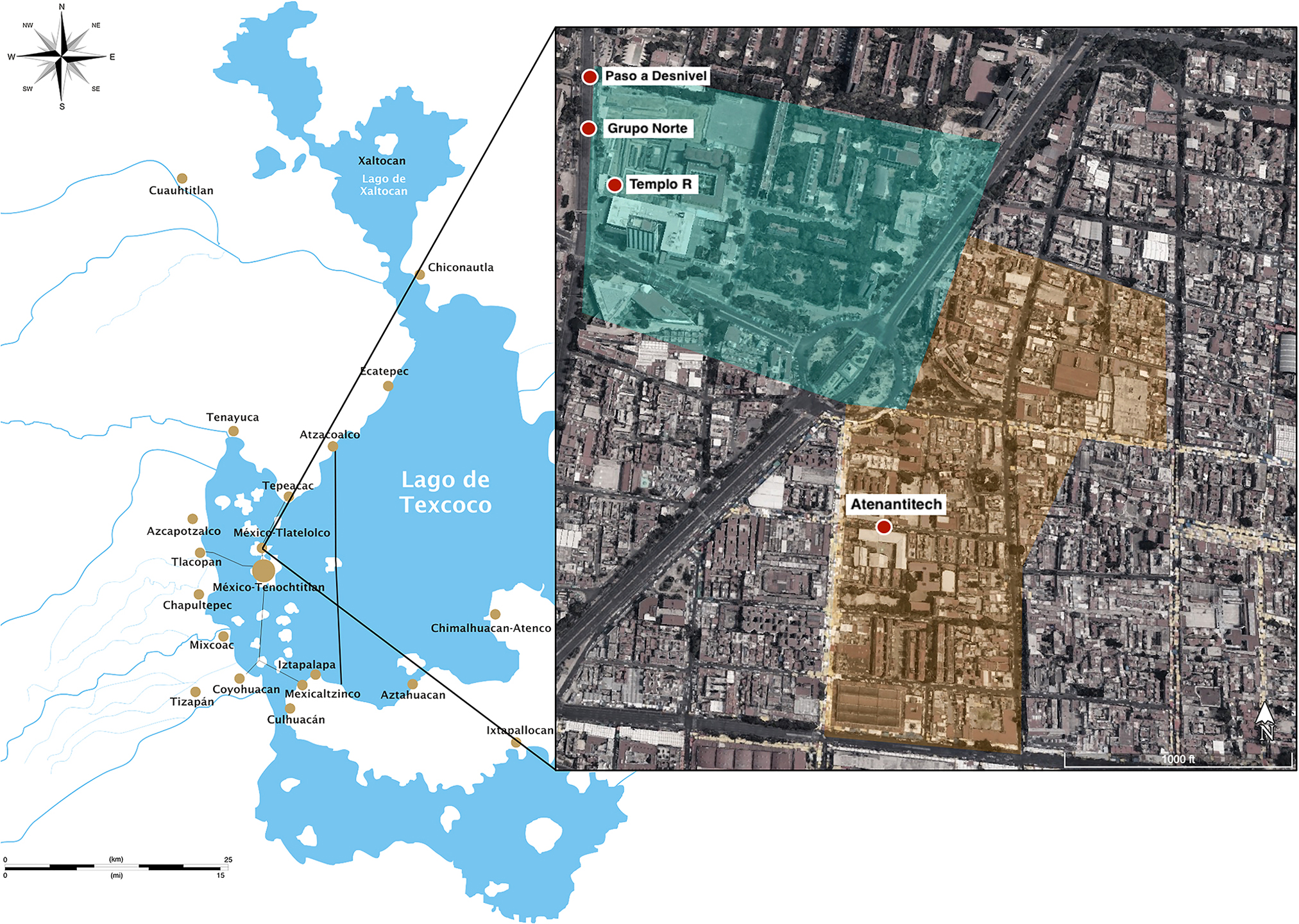
Figure 1. Map of the Basin of Mexico during the Late Postclassic period showing the location of Tenochtitlan-Tlatelolco. Expanded area shows the approximate boundaries of the Tlatelolco ceremonial precinct (teal) and Atenantitech barrio (yellow) overlaid on modern-day Mexico City. Approximate excavation locations of the skeletons considered in this study are marked in red. Basin of Mexico map by Yavidaxiu, public domain, via Wikimedia Commons. Map of Tlatelolco modified from Google, Imagery 2021 CNES/Airbus, Maxar Technologies, following work by HJPD CC BY-SA 3.0.
Materials
Skeletal assemblages analyzed in this study: Grupo Norte, Paso a Desnivel, and Atenantitech
For this study, we analyzed skeletons that were excavated from the Tlatelolco ceremonial center during a period of intense urban development and salvage archaeology in 1961 and 1962. The northwestern area of the site was excavated then, and of particular interest are the areas that presently house the Eje Central Lázaro Cárdenas avenue. A group of structures, Templo I Norte, Templo II Norte, and Templo Redondo, subsequently referred to as Grupo Norte (Fig. 1), had associated burials, previously reported as including primary inhumations, cremations, and mixed burials (González Rul and García Mejía 1962). Hundreds of burials were excavated from Grupo Norte. The iconic Ossuary 14 of more than 150 dismembered, defleshed, and commingled individuals was excavated from this area (Pijoan Aguadé 1997; Pijoan Aguadé et al. 1995), but many single burials were recovered as well. Skeletons were also recovered from the context Paso a Desnivel, which translates to “overpass.” This collection of burials is thought to have been excavated from an area just northwest of Grupo Norte where there now exists a highway overpass on Eje Central Lázaro Cárdenas (Fig. 1).
The primary burials from Grupo Norte (n = 52) and Paso a Desnivel (n = 45) remain unpublished, likely in part due to the aforementioned difficulties delineating mortuary contexts, lack of documented grave goods, and absence of maps, drawings, and levels. As archaeologist Francisco González Rul explained in an interview, the salvage nature of the Grupo Norte and Paso a Desnivel excavations meant that the areas were excavated simultaneously and burials were given numbers as they were excavated, regardless of context (Guilliem Arroyo, personal communication, 2016). For example, Entierros 7–20 and 89–124 were excavated from the Grupo Norte complex, and Entierros 21–71 were excavated from Paso a Desnivel. Thanks to the decades of archival work by Salvador Guilliem Arroyo and the Proyecto Tlatelolco team, it was possible to match the burial numbers and contexts recorded in Francisco González Rul’s field notebook with the burial numbers of the skeletons stored in the Instituto Nacional de Antropología e Historia (INAH) Dirección de Antropología Física (DAF) Tlatelolco collection, stored in the Museo Nacional de Antropología (MNA) in Mexico City, Mexico.
Beginning in 1988, archaeologist Maria de Jesus Sanchez Vazquez led the salvage excavation of a residential area in the neighborhood of Atenantitech, a barrio of Tlatelolco (Caso 1956) (Fig. 1). An area of approximately 5,500 m2 was sampled, and 56 burials were excavated by 1990; 45 of those burials were moved to INAH-DAF for curation, and 40 were available for analysis here (Calderon 2009). Burials were excavated from multiple stratigraphic levels spanning the late Postclassic occupation of Tlatelolco. Although the foundations of residential buildings were found in several levels, all skeletons were excavated from an open area delimited by buildings, further supporting its long-term use as a centro funerario (Jesús Sánchez Vázquez, personal communication). Both adults and non-adults were recovered from the site. Most of the non-adults were buried in a cluster separate from the adults, providing some evidence of age-based mortuary behavior (Calderon 2009). Cremations were apparently excavated from the site as well, but there is no indication of how many. The skeletal assemblage excavated from Atenantitech serves as a normal mortality comparison for the ceremonial center contexts.
Methods
Data collection
The first author collected all the raw age, sex, and pathology data macroscopically from the INAH-DAF Tlatelolco skeletal collections at the MNA in Mexico City, Mexico.
Sex recording details
Sex was primarily assessed using pelvic morphology. When necessary due to incomplete preservation, however, cranial morphology and scapula glenoid, humeral head, and/or femoral head width measurements were used to assign sex. No attempt was made to assess the sex of non-adult skeletal morphology (Appendix S1 Methodology: Sex assessment).
While reconciling context and burial numbers as recorded in the original field notebook with burial numbers on the storage boxes, however, it was possible to match skeleton IDs to those published as part of aDNA studies. Therefore, genetic sex assignments were included for the Grupo Norte and Paso a Desnivel individuals analyzed by Morales-Arce et al. (2019) and for individuals in the comparative Templo R sample analyzed by De La Cruz et al. (2008) (Table A1).
Age recording details
Non-adults. When teeth were available, age range estimates were generated using The London Atlas of Tooth Development and Eruption (AlQahtani et al. 2010). Otherwise, age was estimated using skeletal element measurements (Maresh 1970) and/or epiphyseal union stages as collated by Schaefer et al. (2009) (see Appendix S1 Methodology: Non-adult age assessment). For statistical treatment, the average value of each non-adult’s age range was selected as a point estimate for all summary statistics and analyses.
Adults. Age ranges were generated using Transition Analysis ADBOU Age Estimation v2.1.046 (available at http://statsmachine.net/software/ADBOU2/) for individuals whose epiphyses were obliterated and dental development was complete (Boldsen et al. 2002). The corrected point estimate was rounded to the nearest whole number and used as the age estimate for all summary statistics and analyses. Age ranges and point estimates for all individuals can be found in Table A1.
Paleopathological recording details
Differential diagnosis
A differential diagnosis was designed using published criteria for scurvy, rickets, porotic lesions caused by acquired anemia, and infectious disease (Klaus and Lynnerup 2019; Ortner and Mays 1998; Ragsdale et al. 1981; Schattmann et al. 2016; Snoddy et al. 2018; Stuart‐Macadam 1991; Weston 2012). Each individual was assigned present, absent, or unobservable for each pathological indicator. See Appendix S2 (A, B, and C): “Pathology Distribution Figures” for intra-skeleton preservation and pathological distributions. Diagnostic pathological changes caused by rickets were not observed in any individuals, so the final pathological categories for the analysis were scurvy, anemia, and infectious disease (Table A2). See Appendix S1 Table S1 missing data assessment for a summary of how unobservable/missing values affected sample sizes for each analysis.
Scurvy. Individuals were diagnosed with scurvy if they had at least one diagnostic indicator or at least two suggestive indicators as defined in Table A2. The differential diagnosis was designed using criteria published by Snoddy et al. (2018) and Schattmann et al. (2016), and photographic examples of indicators can be seen in Figure 2. Individuals were determined to be scurvy free if they had cranial and postcranial elements preserved and no diagnostic lesions or only one suggestive lesion. In this study, most scorbutic changes were isolated to cranial and mandibular elements. Therefore, if an individual did not have a preserved skull or cranial fragments and had no postcranial pathological changes diagnostic or suggestive of scurvy, they were identified as unobservable (NA) for scurvy.
Anemia. Penetrating porotic lesions accompanied by expansive diploë and thinning of the outer table on the orbits and cranial vault were considered indicative of anemia (Types 3, 4, and 5 from Stuart-Macadam 1991). If an individual did not have a preserved skull or cranial fragments, they were identified as unobservable (NA) for anemia.
Infectious disease. An individual was identified as having infectious disease if they had subperiosteal new bone formation (SPNBF), osteolytic changes, or osteoblastic changes suggestive of infection. In this study, diffuse SPNBF was recorded as infection due to its documented and hypothesized associations with systemic bacterial and viral infections (Burrows 1971; Csonka and Pace 1985; Haygood and Williamson 1994; Lakey et al. 2008; Rasool 2001; Teo and Peh 2004). SPNBF was identified as an indicator of infection when the lesion patterning suggested a systemic stimulus (Ragsdale et al. 1981; Weston 2012). Specifically, the SPNBF must have been (1) present on at least a third of the diaphysis of long bones and (2) bilaterally distributed or present across multiple skeletal elements. Additionally, individuals with SPNBF along the visceral surface of the vertebral portion of the ribs were identified as having an infection (Cheng Pau et al. 2009; Collier et al. 1967; Davies-Barrett et al. 2019; Roberts et al. 1994). SPNBF lesions formed in response to the pooling of blood and subsequent inflammation on the distal aspects of long bones, thought to be caused by scorbutic hemorrhaging (Choi et al. 2007; Gulko et al. 2015), were not considered as indicators of infection.
In several cases, osteolytic and osteoblastic changes could be reliably attributed to a more specific disease process, such as treponemal disease and tuberculosis (Baker et al. 2020; Klaus and Lynnerup 2019; Pálfi et al. 2012). Those data will be analyzed in subsequent publications, but in this analysis, those individuals were recorded as having infectious disease. Therefore, “infectious disease,” as recorded here, refers to unspecified and specific infections. Only individuals with more than half of their appendicular and axial skeletons preserved were recorded as observable for infectious disease.
Comparative age and sex data. The well-documented sacrificial assemblage from Templo R was used as a comparative sample for paleodemographic analyses. These skeletons are curated in the Tenochtitlan Templo Mayor Museum and were not analyzed as part of this study, so they are not included in the pathology analyses. We used age estimates previously published by Moreiras Reynaga et al. (2021), De la Cruz et al. (2008), and Guilliem Arroyo (1999), as indicated in Table A1.
Cut marks. The first author documented perimortem cut marks typical of those previously identified at Tlatelolco by Pijoan Aguadé (1995, 1997). All skeletal elements were macroscopically examined for perimortem trauma. Perimortem trauma related to ritual treatment was classified as dismemberment, scalping, cut marks from defleshing and/or disarticulation, heart extraction, or decapitation (following Hamilton 2016; Jelíneck 1993; Pijoan Aguadé 1997; Pijoan Aguadé and Mansilla Lory 2010; Pijoan Aguadé et al. 1995; Tiesler and Olivier 2020). Dismemberment was identified as percussive impacts near ligament attachments or joints (Fig. 3a). Scalping was identified as cut marks on the calvarium (Fig. 3b). Defleshing/disarticulation was identified as clusters of cut marks around tendon or muscle attachment sites, including the vertebral rib ends (Fig. 3c). Heart extraction was identified as cut marks on the sternal ends of the ribs, multiple perimortem fractures on sternal rib ends (Fig. 4a, b), and/or bisected sterna and/or manubriam (Fig. 4c). Decapitation was identified as cut marks on the basicranium, cervical vertebrae, and/or first rib. Using written descriptions and photos, composite distributions of perimortem trauma were created in Pixelmator Pro v2.0.5 Junipero.
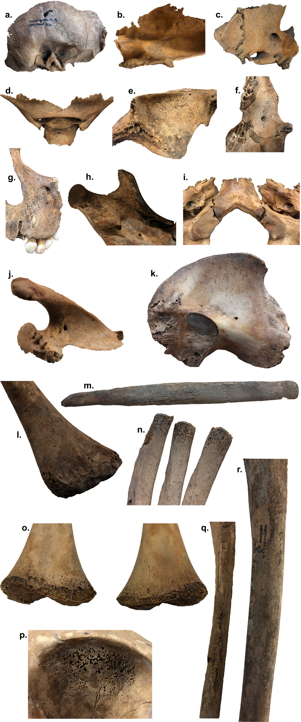
Figure 2. Examples of pathological indicators used in the differential diagnosis (skeleton ID). (a) Ectocranial temporal and greater sphenoid wing porosity and new bone formation (NBF) (196_116). (b) Endocranial occipital NBF (128_40). (c) Endocranial sphenoid foramen rotundum NBF (128_40). (d) Endocranial sphenoid lesser wing NBF (121_24). (e) Orbit NBF and porosity (118_19). (f) Posterior zygomatic and posterior maxilla NBF (195_114). (g) Anterior maxilla and infraorbital foramen porosity (195_114). (h) Medial mandible coronoid process porosity (118_19). (i) Inferior pars basilaris porosity (56_14D). (j) Scapula supraspinous fossa NBF and porosity (121_24). (k) Visceral surface ilium NBF, vascular impressions, and porosity (195_114). (l) Distal anterolateral humerus metaphysis NBF and porosity (121_24). (m) Anterolateral rib shaft NBF and porosity (182_94). (n) Rib sternal end porosity (183_95b). (o) Anterior distal femur metaphysis porosity (56_14D). (p) Orbit penetrating and expansive porosity (ATN_22_28). (q) Anterior ulnae diffuse NBF (121_24). (r) Anterolateral tibia diaphysis NBF (129_43). Photos taken by first author or Juan Salvador Rivera Sánchez INAH-DAF (h).
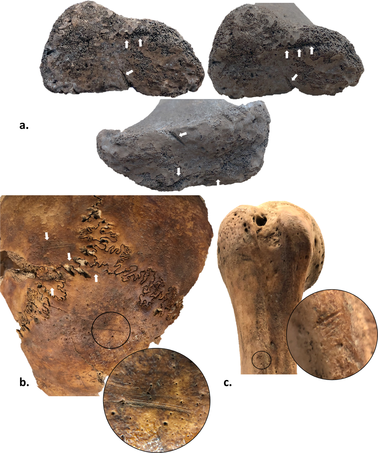
Figure 3. (a) Right femur distal epiphyseal surface, three angles; impact from dismemberment; Grupo Norte 182_94. (b) Posterior parietals and occipital; scalping with inset; Paso a Desnivel 135_55. (c) Right proximal humerus; cut marks (defleshing and/or disarticulation) on crest of greater tubercle with inset; Paso a Desnivel 137_59.
Radiocarbon dating
To understand better the chronology of the mortuary deposits, seven individuals were chosen for radiocarbon dating: three from Grupo Norte, three from Paso a Desnivel, and one from Atenantitech. Samples of six vertebral elements and one ilium weighing between 344 and 693 mg were processed by the University of Arizona AMS Laboratory.
Comparative analyses
Paleodemographic comparisons
To differentiate between special mortuary and normal mortality deposits, we compared the age-at-death distributions of the three ceremonial center contexts, Grupo Norte, Paso a Desnivel, and Templo R, and one residential context, Atenantitech. The distributions were visualized by binned age categories as well as by Kaplan–Meier survival curves. To determine if there are significant differences among the mortuary contexts, two multiway (1: all contexts; 2: ceremonial center contexts) and one pairwise (residential context and ceremonial center context) log-rank tests were performed on the survival times (i.e., age-at-death distributions). Log-rank test was chosen because we are only interested in differences in overall survival curve distribution and not controlling for any confounding factors. To correct for the family-wise error rate that results from performing multiple statistical tests, the Bonferroni adjustment was made by dividing the original alpha value by the number of log-rank tests, for a corrected alpha value of α = 0.05/3 = 0.016.
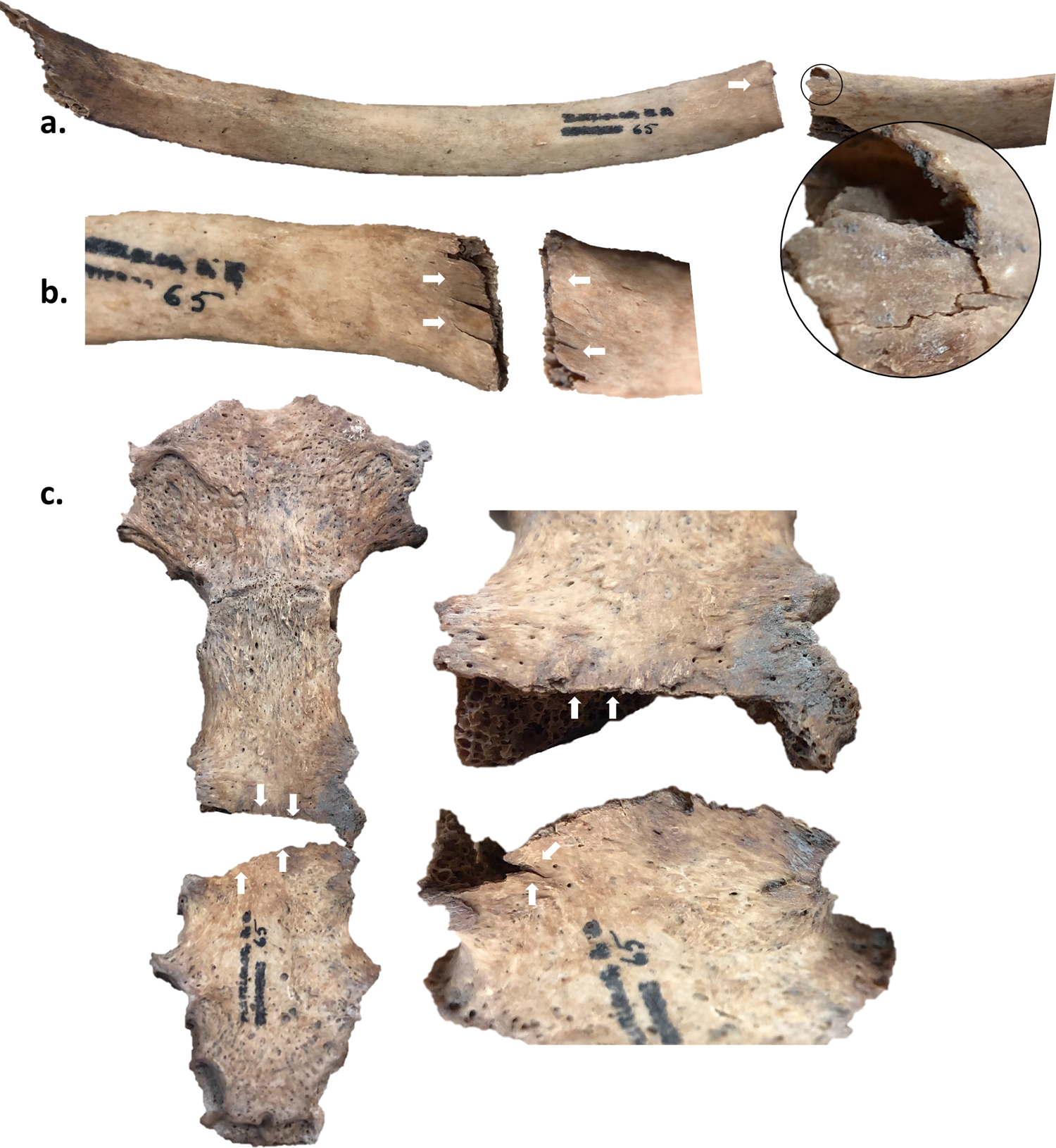
Figure 4. Paso a Desnivel 140_65. (a) Left rib #4 visceral surface showing perimortem break on sternal end (left) and same rib with view of external surface showing perimortem break (right) with black circle showing plastic deformation. (b) Right rib #2 showing perimortem break at the sternal end from the visceral (left) and external (right) surfaces. (c) Perimortem bisection of sternum. White arrows showing lack of color differential and/or plastic deformation.
In addition to age-at-death distributions, we compared sex ratios across all contexts. Sex of individuals younger than 16 years was previously assessed using amplicon (De la Cruz et al. 2008) and whole-genome sequencing (Morales-Arce et al. 2019).
Metabolic and infectious disease distributions
Chi-square analysis. First, we performed a chi-square test on a 3 × 4 contingency table to identify differences in metabolic disease, infectious disease, and comorbidity of infectious and metabolic disease distributions between the ceremonial center contexts (Grupo Norte and Paso a Desnivel) and residential context (Atenantitech). Scurvy and anemia were collapsed into a metabolic disease category to avoid uncertainty in the etiology of orbital lesions. To ensure comparable results across contexts, analyses were limited to individuals with age estimates younger than 20 years because (1) the ratio of non-adults to adults is higher in the ceremonial center contexts, and (2) skeletal manifestations of pathologies differ between non-adults and adults. The cutoff of 20 years was chosen to include adolescent individuals with non-obliterated epiphyses who had age ranges spanning 16 to 22 years. Although it is recommended to use a Fisher’s exact test for contingency tables when >20 percent of the cells have values fewer than five, a chi-square test was chosen because it is better suited for the interpretation and visualization of multiway contingency tables (SI Methodology: Chi-square visualizations). To be conservative, we performed Fisher’s exact tests and found similar p-values for both tests.
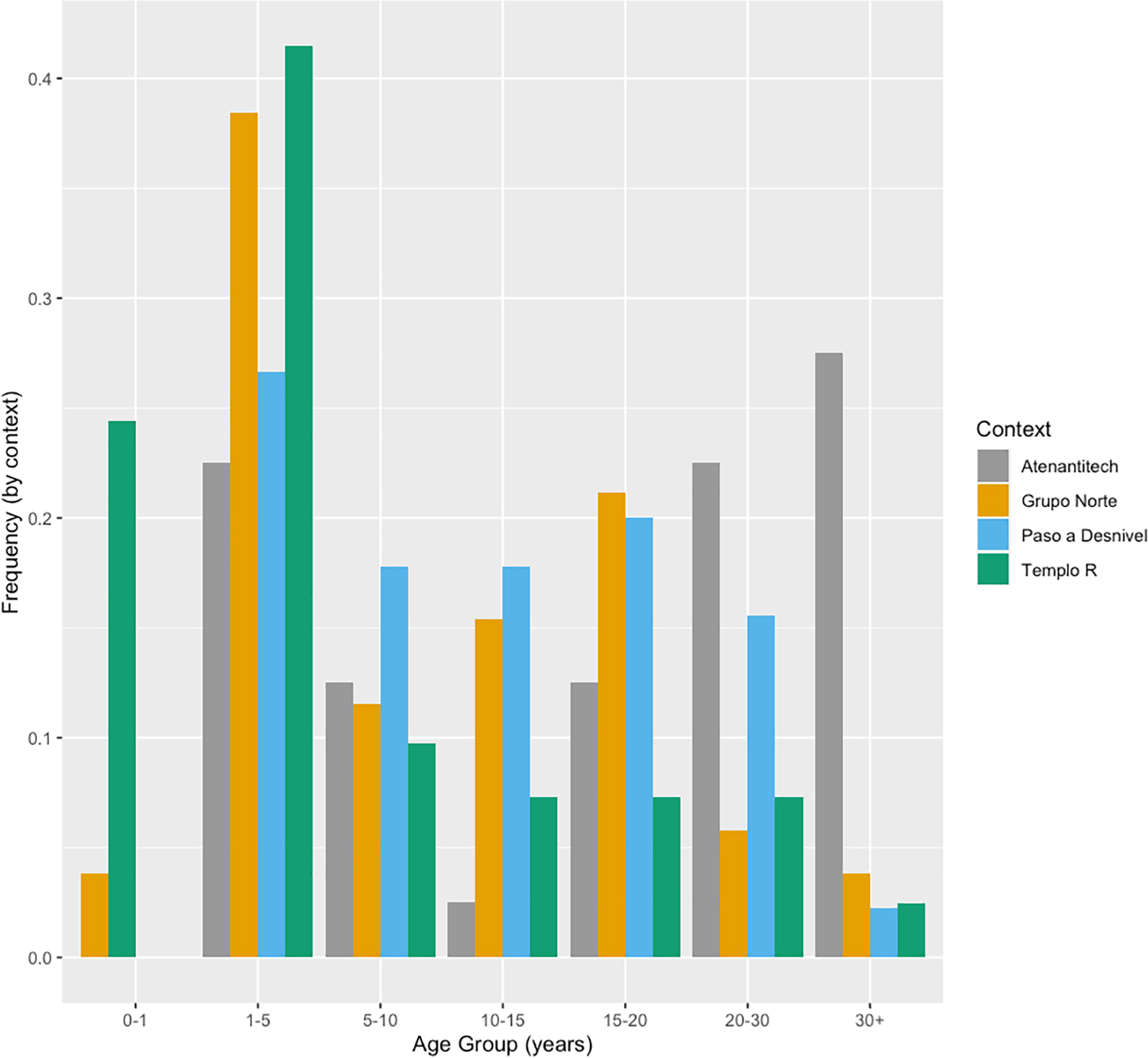
Figure 5. Age-at-death distribution by context. For the age ranges, the lower value is exclusive, and the higher value is inclusive. For example, Age Group 1–5 contains individuals more than one year of age at death up to and including five years of age at death, whereas individuals one year or less of age at death are in Age Group 0–1.
All statistical tests and plotting were done in RStudio (R version 4.1.2) using base R and the ggplot2, ggridges, Bchron, tidyr, dplyr, tibble, survminer, survival, and corrplot packages (Haslett and Parnell 2008; Kassambara et al. 2019; Müller and Wickham 2020; Therneau 2015; Wei and Simko 2017; Wickham 2016, 2020; Wickham et al. 2020; Wilke 2017). All of the R code can be found at https://github.com/Kelzor/Human-sacrifice-and-malnutrition-at-Tlatelolco.
Cut marks. The perimortem trauma distributions were visually compared using the composite distributions and a bar chart of counts.
Results
Paleodemographic comparisons
There are clear differences in age-at-death distributions between ceremonial and residential contexts (Fig. 5). The residential context, Atenantitech, approximates a normal-mortality distribution as a U-shaped curve with peaks in the first five years of life and during adulthood. There were no perinates or infants recovered from the Atenantitech cemetery, suggesting perinates and infants had distinct mortuary treatment. In contrast to the U-shaped distribution of Atenantitech, the Grupo Norte and Paso a Desnivel distributions are dominated by peaks in the 1- to 5-year and 10- to 20-year age categories and have few adult individuals. The Templo R distribution differs from all other contexts with the largest peaks in the youngest age categories, 0–1 and 1–5.
Kaplan–Meier survival curves illustrate the probability of individuals remaining in the sample as age increases (Fig. 6). The log-rank test result for all contexts indicates that there is a significant difference among survival curves (p < 0.001) (Fig. 6a, Table 1). There are significant differences among survival curves of the ceremonial center contexts (p = 0.007) (Fig. 6b) and between the residential context, Atenantitech, and the ceremonial center context with the most similar survival curve, Paso a Desnivel (p = 0.004) (Fig. 6c, Table 1). The age-at-death distributions of Atenantitech and Templo R are significantly different from one another and the other two ceremonial center contexts, Grupo Norte and Paso a Desnivel.
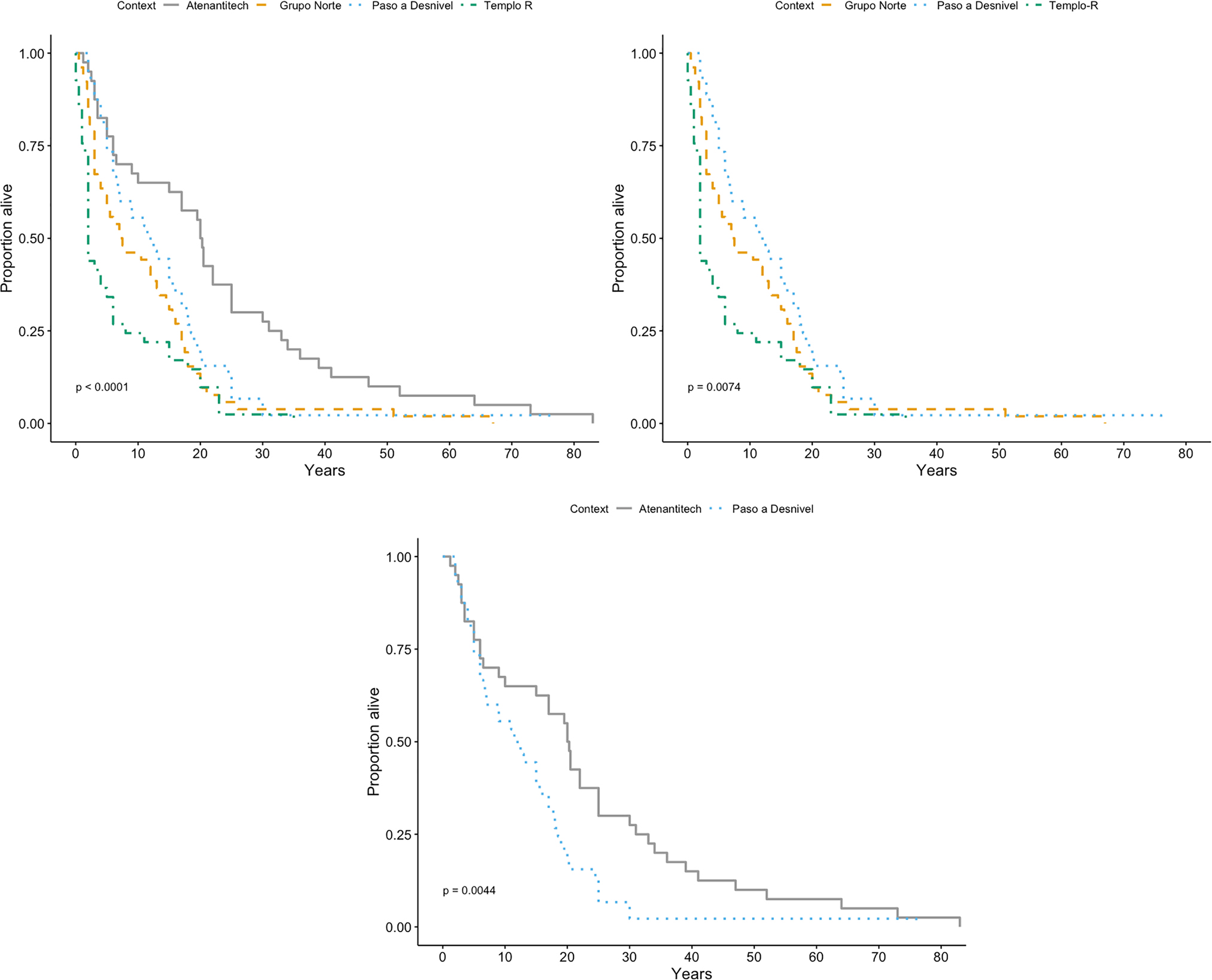
Figure 6. Survival curves of (a) all contexts, (b) ceremonial center contexts, and (c) residential and most closely related ceremonial center context.
Table 1. Log-Rank Test Results by Context Comparison.
|
Comparison |
χ2 |
df |
p |
|||
|
All contexts |
32.4 |
3 |
<0.001* |
|||
|
Ceremonial contexts |
9.8 |
2 |
0.007* |
|||
|
Residential and most similar ceremonial |
8.1 |
1 |
0.004* |
*Significant at α = .05/3 = .016.
There is a marked sex skew in non-adults from the ceremonial center (Fig. 7). The Templo R non-adults are almost entirely male, and the Paso a Desnivel and Grupo Norte non-adults are almost entirely female. The individuals aged 16+ years-at-death from Templo R are all male. The 16+ individuals from Paso a Desnivel and Grupo Norte show less sex bias than in the younger individuals, but the older individuals are predominantly female. No individuals younger than 16 years from Atenantitech have sex assessments because biomolecular work has not been performed on these remains. The 16+ individuals from Atenantitech approximate an even female to male ratio at 56:44.
Metabolic and infectious disease distributions
The comparison of infection, metabolic disease, and comorbidity distributions across the three contexts was limited to individuals younger than 20, because pathology frequencies are similar within contexts and age groups before 20+ years (Fig. 8). There are clear visual differences in pathology prevalence among contexts (Fig. 9). The distributions of pathology across all contexts are significantly different, χ2(6, n = 85) = 25.812, p < 0.001. The distributions of pathology between the two ceremonial center contexts are significantly different as well, χ2(3, n = 71) = 11.441, p = 0.009. The pathology distributions between Atenantitech and Grupo Norte do not significantly differ, χ2(3, n = 51) = 7.427, p = 0.059 (Table 2) (see Appendix S1 Figure S1 Chi-square visualizations).
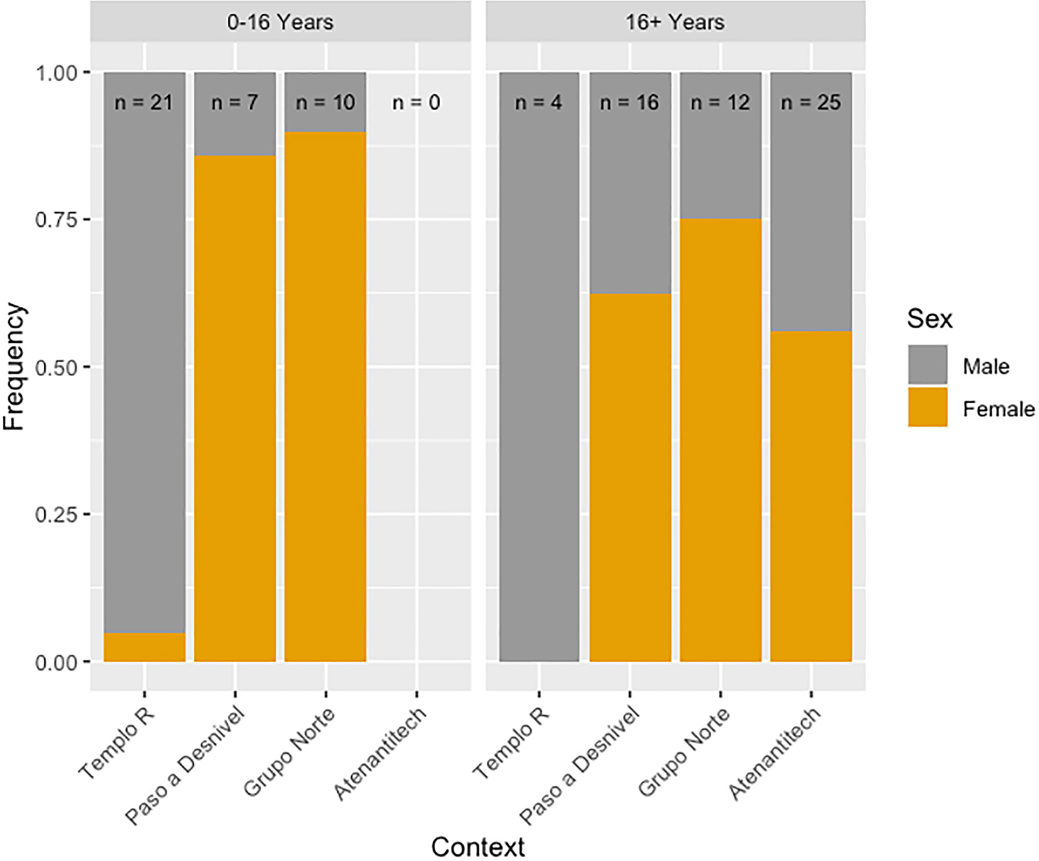
Figure 7. Sex prevalence by context and age category. Sample sizes indicate the number of individuals for which sex assessments were possible. Genetic sex determination of individuals in the 0–16 age category was performed by De La Cruz et al. (2008) (Templo R) and Morales-Arce et al. (2019) (Grupo Norte and Paso a Desnivel).
The pathology distribution by age category and context reveals that the percentage of unaffected individuals from Atenantitech remains similar across all age categories, despite small sample sizes per age category (Fig. 8). Individuals from the ceremonial center deposits show evidence of infection co-occurring consistently with metabolic disease in non-adults 0–10 years of age-at-death. Infectious and metabolic comorbidity, however, does not occur in the youngest individuals from Atenantitech. All the individuals aged 20+ from the ceremonial center deposits are affected. Among the Atenantitech age groups, the individuals aged 20+ have the highest prevalence of infection without co-occurrence of metabolic disease.
Cut marks
Paso a Desnivel has five to six times as many occurrences of perimortem trauma as the other two contexts (n = 21), and it is the only context with evidence of heart extraction (Fig. 10, Table 3). Grupo Norte (n = 4) and Atenantitech (n = 3) have similar amounts of perimortem trauma, but the types of ritualized traumas differ between the two sites (Figs. 10 and 11, Table 3). The only forms of ceremonial perimortem trauma identified at Atenantitech are defleshing/disarticulation and scalping, whereas dismemberment is also present at Grupo Norte. All types of perimortem trauma were observed across all age groups, except for heart extraction, which was only observed in two adults, a female and male. There are no apparent sex-based differences among perimortem traumas, but only 60 percent of individuals have sex assignments (Table 3).
Radiocarbon dating
All seven bone samples yielded sufficient collagen for analysis, and the atomic C/N ratios range from 3.2 to 3.4, which is considered to be of sufficient quality (Deniro 1985). The IntCal20 (Reimer et al. 2020) calibrated dates from the Grupo Norte samples, two Paso a Desnivel samples and the sample from Atenantitech, were dated between 1332 and 1445 CE (95 percent probability interval) (Fig. 12, raw data in Appendix S1 Table S2). One sample from Paso a Desnivel was dated to 897–1025 CE (95 percent probability interval).
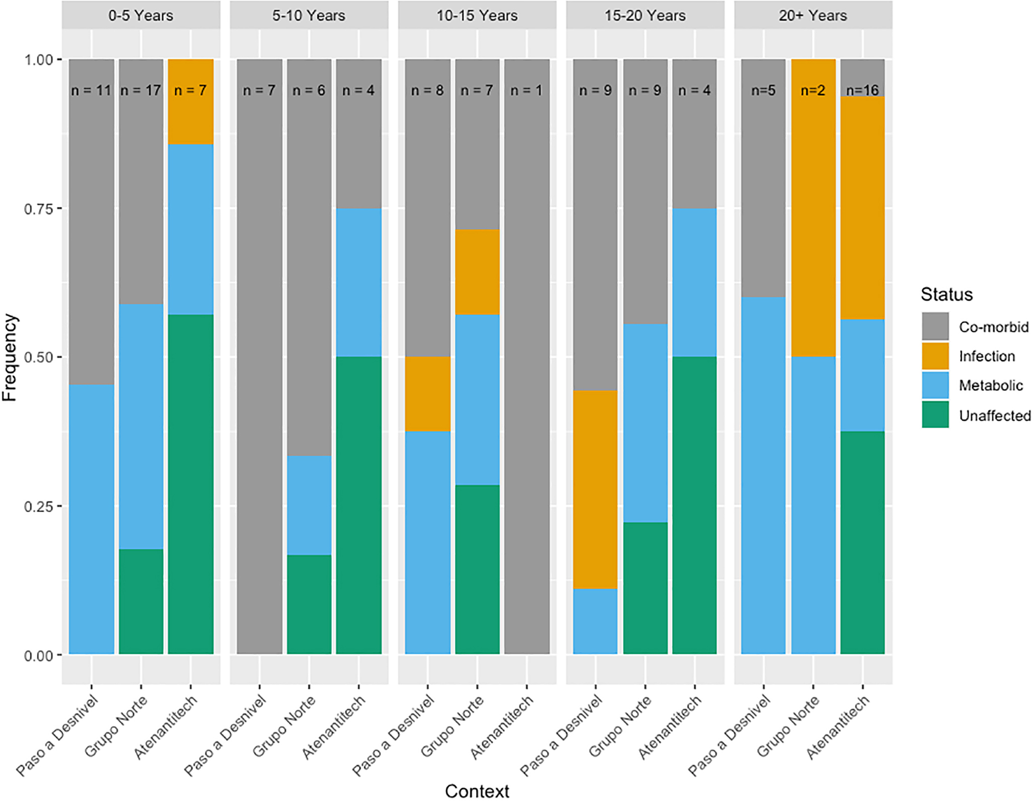
Figure 8. Prevalence of pathology by context across age groups. Sample size is indicated for each context for each age category. Note that there is only one observable individual from Atenantitech for the 10–15 year age category.
Were the Individuals from the Ceremonial Center Sacrificed?
Despite the limitations of analyzing skeletons with incomplete provenience information, we were able to identify meaningful trends within and among contexts that strongly suggest at least some, if not all, of the ceremonial center individuals were ritually killed; individuals buried in the ceremonial center deposits are significantly younger with skewed sex ratios, have significantly more evidence of metabolic and infectious disease, and were ritually processed with greater variability than those buried in the residential context of Atenantitech.
Given that the ethnohistorical and (bio)archaeological records indicate that the ritual killing of children was an integral part of Mexica religion and enabled dialogue between the earthly and supernatural realms, it is unsurprising that there would be many non-adult sacrificial deposits at Tlatelolco. While the residential context of Atenantitech approximates a normal mortality distribution, the age distributions of Grupo Norte, Paso a Desnivel, and Templo R are significantly younger (Fig. 3), with high frequencies occurring in the 15–20 age group, when individuals should be at their most resilient (Weiss 1973). The Grupo Norte and Paso a Desnivel age distributions deviate from that of Templo R (Fig. 5), with higher frequencies of older children (5–10 and 10–15) and adolescents (15–20) (Fig. 3). Moreover, the Grupo Norte and Paso a Desnivel non-adults who have genetic sex assessments are predominantly female, the opposite of what is observed at Templo R (Fig. 7). The skew toward female inclusion is also present in the adult individuals, but it is less pronounced. It is possible that these deposits are combinations of sacrificial victims, war captives, slaves, and children, which has been described by colonial-period chroniclers (Horcasitas and Heyden 1971:256), chosen for their embodiment of a female deity. Compared to Templo R, Grupo Norte and Paso a Desnivel have a marked absence of infants (defined here as 0–1 year), which could reflect the different roles played by these victims of ritual killings. Similar to observations by Román Berrelleza (1990, 1999, and 2010), the individuals from the ceremonial center contexts are characterized by a high frequency of porotic cranial and orbital lesions. In the case of Paso a Desnivel, all individuals have evidence of metabolic disease, infection, or both. Although the difference in pathology distribution between Grupo Norte and Atenantitech is not statistically significant, Atenantitech has the highest frequency of unaffected non-adults and the lowest frequency of comorbidities overall (Fig. 9).
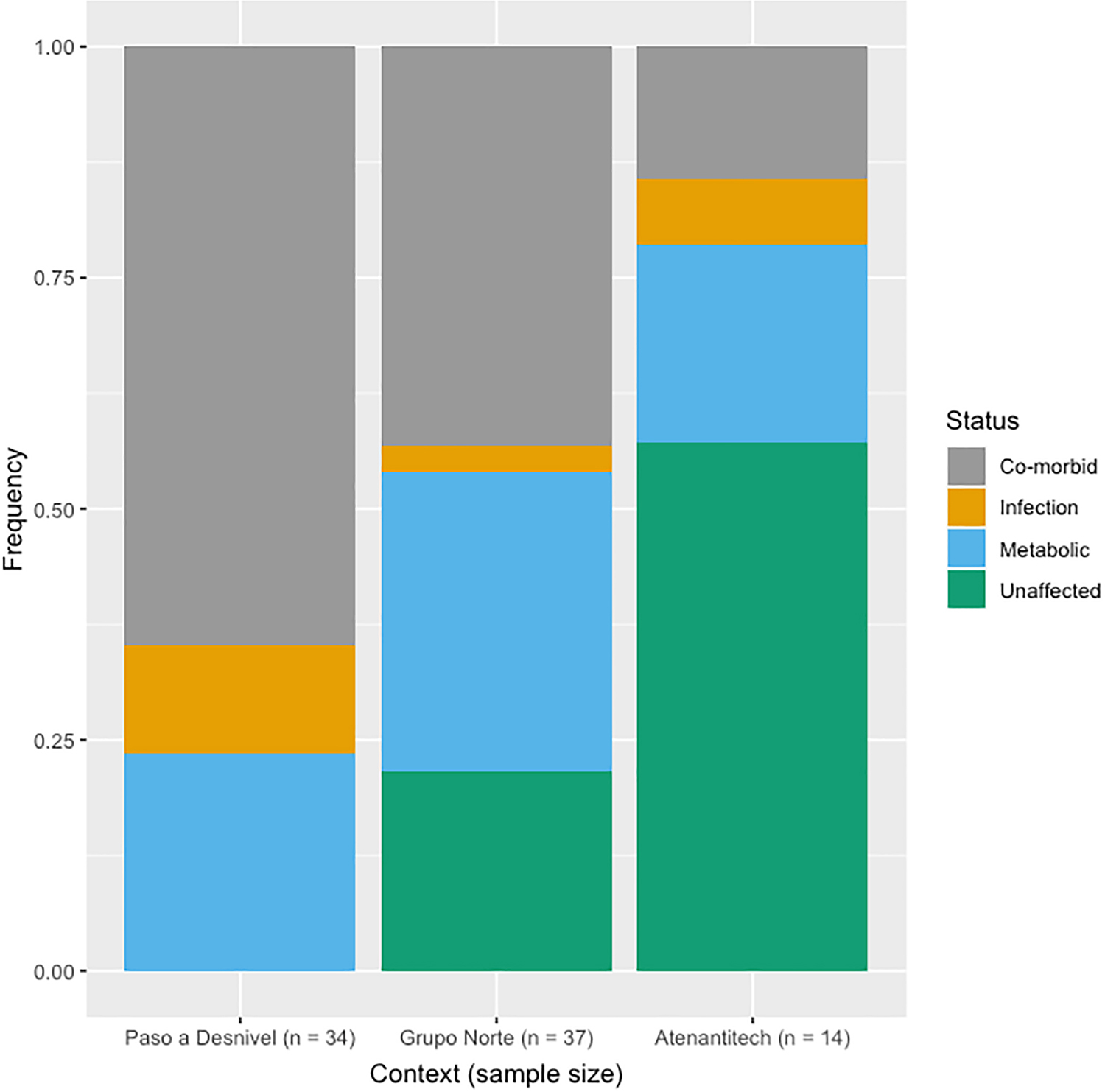
Figure 9. Prevalence of pathology in individuals younger than 20 years by context.
Table 2. Contingency Tables and Chi-Square Test Results for Each Combination of Contexts.
|
Comorbidity |
Infection |
Metabolic |
Unaffected |
|||||
|
Paso a Desnivel |
22 |
4 |
8 |
0 |
||||
|
Grupo Norte |
16 |
1 |
12 |
8 |
||||
|
Atenantitech |
2 |
1 |
3 |
8 |
||||
|
χ2 |
df |
N |
p |
|||||
|
25.81 |
6 |
85 |
<0.001* |
|||||
|
Comorbidity |
Infection |
Metabolic |
Unaffected |
|||||
|
Paso a Desnivel |
22 |
4 |
8 |
0 |
||||
|
Grupo Norte |
16 |
1 |
12 |
8 |
||||
|
χ2 |
df |
N |
p |
|||||
|
11.44 |
3 |
71 |
0.009* |
|||||
|
Comorbidity |
Infection |
Metabolic |
Unaffected |
|||||
|
Grupo Norte |
16 |
1 |
12 |
8 |
||||
|
Atenantitech |
2 |
1 |
3 |
8 |
||||
|
χ2 |
df |
N |
p |
|||||
|
7.42 |
3 |
51 |
0.059 |
*Significant at α = .05/3 = .016.
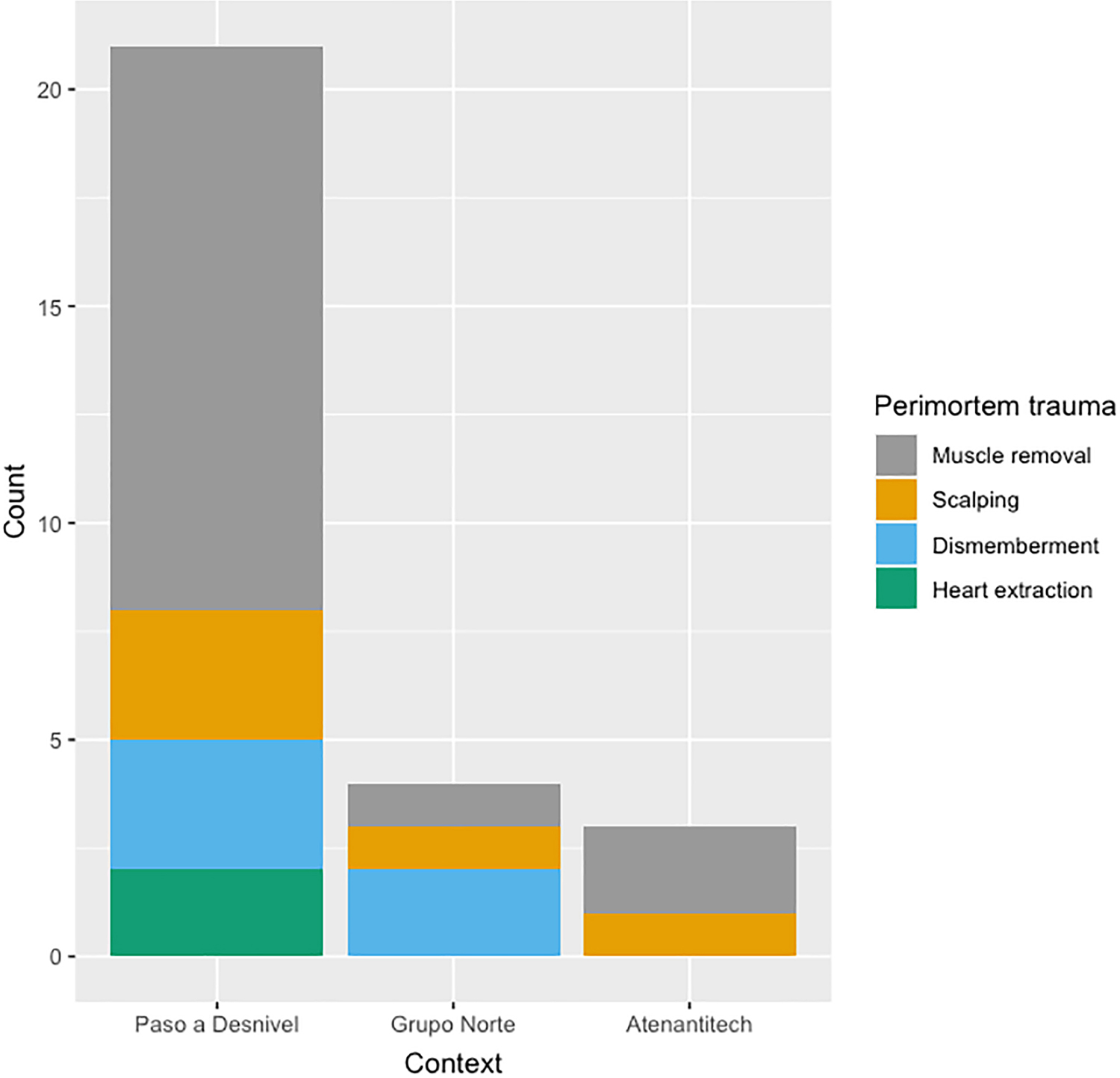
Figure 10. Count of each type of perimortem trauma by context. Some individuals are represented more than once because they had more than one type of peri-postmortem trauma.
Table 3. Perimortem trauma occurrence by context, age, and sex. Sex assignments of non-adults were made from genetic X and Y chromosome data by Morales Arce et al. (2019).
|
Context |
Perimortem Trauma |
Number of Individuals |
Age-at-Death Point Estimate in Years (Sex Where Applicable) |
|||
|
Paso a Desnivel |
Defleshing/disarticulation |
8 |
4, 5, 5, 6 (F), 9, 15 (F), 15, 20 (F) |
|||
|
Defleshing/disarticulation and scalping |
1 |
18 (M) |
||||
|
Defleshing/disarticulation and dismemberment |
3 |
18.5 (M), 19.5 (F), 20.25 (M) |
||||
|
Defleshing/disarticulation and heart extraction |
1 |
24.5 (F) |
||||
|
Scalping |
1 |
11.5 (M) |
||||
|
Scalping and heart extraction |
1 |
30 (M) |
||||
|
Grupo Norte |
Defleshing/disarticulation |
1 |
0.75 |
|||
|
Scalping |
1 |
2.5 |
||||
|
Dismemberment |
2 |
4.75 (F), 11.5 |
||||
|
Atenantitech |
Defleshing/disarticulation |
2 |
17 (F), 25 (M) |
|||
|
Scalping |
1 |
3.5 |
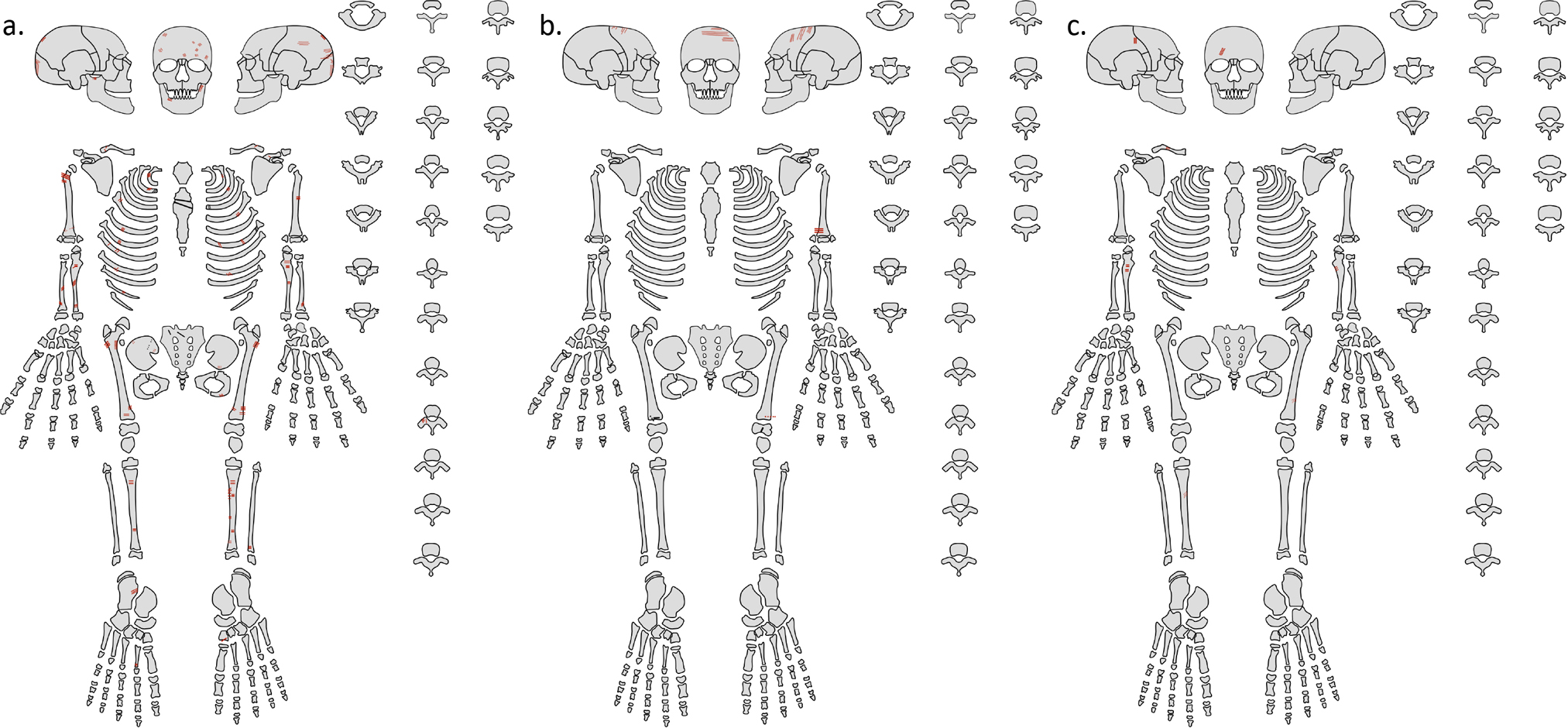
Figure 11. Composite perimortem trauma distribution for (a) Paso a Desnivel, (b) Grupo Norte, and (c) Atenantitech. Red indicates shallow cut marks. Black indicates percussive impacts and perimortem fractures. A dotted line indicates the perimortem trauma is on an aspect of the skeleton not visible (e.g. posterior, inferior, or superior).
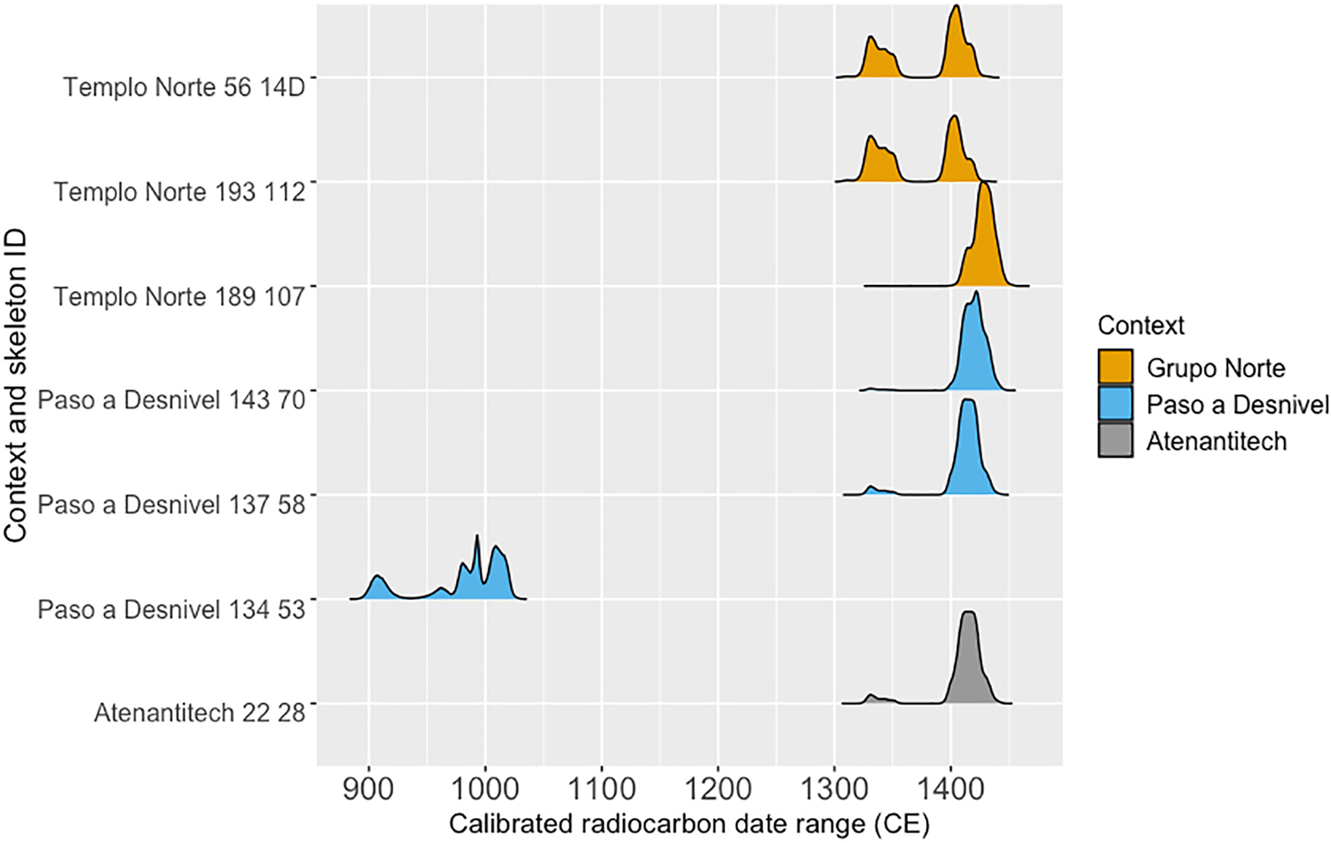
Figure 12. Ridgeline plot of radiocarbon dating probability distributions colored by context.
Given that evidence for some perimortem trauma was identified in all contexts analyzed here, it is surprising that no such evidence has been reported from Templo R or Ofrenda 48, aside from a single bisected femur (Román Berrelleza 1990). The reported method of child sacrifice involved throat cutting, which would not necessarily leave skeletal evidence. Indeed, Medrano Enríquez (2021) also remarks on the lack of perimortem trauma from throat cutting, cut marks on the cervical vertebrae or basicranium, in sacrificed non-adults from Tula (1000–1200 CE). Except for the two cases of heart extraction from Paso a Desnivel, the perimortem trauma likely reflects distinctive ritualized treatment for the bones and flesh after death. Ethnohistoric accounts describe an intimate relationship between sacrifice and ceremonial consumption by nobles, priests, and warriors (Anderson and Dibble 1981:24, 29; Carrasco 1999:84, 174–176; Heyden 1994:192–193; Horcasitas and Heyden 1971:191). This ceremonial consumption of sacrificed flesh, along with the ritualized flaying and re-purposing of skins (Carrasco 1999:145; Heyden 1994:485), could explain the evidence for soft tissue removal. Further, dismembered body parts were deposited in funerary spaces as a way to consecrate the ground (Núñez Enríquez 2006:149), but it is possible that other sacrificial and ceremonial acts could have been performed to sanctify burials. This could explain the evidence for soft tissue removal at Atenantitech. Interestingly, the context with the highest frequency of pathology is also the context with the most counts of perimortem trauma (Paso a Desnivel).
Together, the demographic homogeneity, high pathology prevalence, and perimortem trauma strongly suggest that Grupo Norte and Paso a Desivel, like Templo R and Ofrenda 48, were sacrificial deposits. Further, interpreted within the osteological paradox, the trend of high comorbidity and low unaffected prevalence within these contexts suggests that these individuals were not dying natural deaths (Wood et al. 1992). That so many of these individuals have skeletal indicators of infectious and metabolic disease suggests that they were robust to have survived until the point of sacrifice. Some of these individuals likely could have survived into adulthood.
There are few comparative data for sacrificial deposits of single burials from Mexica sites, as much of the mortuary research has focused on ossuary deposits with clear evidence of sacrifice, elite burials, or caches of decapitated crania (Chávez Balderas 2017; González Rul 1997; Pijoan Aguadé 1997;Pijoan Aguadé et al. 1989; Pijoan Aguadé and Mansilla Lory 1997, 2010). There are, however, some comparable examples from Central Mexico. Within Central Mexico, there is only one example of a middle-late Postclassic non-adult mass burial, to the authors’ knowledge. At Teopanzolco, Morelos, approximately 100 km south of Tenochtitlan-Tlatelolco, a cache of at least 92 individuals was dated to the middle-late Postclassic period using ceramic burial goods (Lagunas Rodríguez and Serrano Sanchez 1972; Smith 2010). Over half of the individuals were non-adults, and most individuals had skeletal evidence of disarticulation or limb, hand, or foot removal. Of the adults, males and females were represented equally (Lagunas Rodríguez and Serrano Sanchez 1972). Two Postclassic burials from Cholula, Puebla, demonstrate small-scale child sacrifice, more like accounts by Spanish chroniclers than the larger deposits at Teopanzolco and Tenochtitlan-Tlatelolco. Two children were excavated from a central altar within the ceremonial plaza; their skulls were found disarticulated and arranged away from the postcranial elements (Lagunas Rodríguez et al. 1976:74). Gabriel de Rojas (a colonial mayor of Cholula) reported in 1581 that human sacrifices were performed in Cholula during times of drought. He described the sacrifice of children captured or bought of ages 6–10 (Rojas 1927).
Due to the absence of maps, levels, drawings, and original burial photos, we assessed burial chronology through radiocarbon dating. Six out of seven date estimates fall between 1332 and 1445 CE (95 percent probability interval), in agreement with the generally accepted timeframe for the founding of Tenochtitlan and Tlatelolco and the subsequent growth in size and influence of the connected cities. Interestingly, one individual from Paso a Desnivel, 134-53, was dated to 897–1025 CE (95 percent probability interval), approximately 300–400 years before the supposed founding of the cities. The earlier date from Paso a Desnivel suggests that at least this area of the site and probably others were recurrently used for sacrificial deposits for centuries. It is notable that the individual dated 300–400 years earlier than the others, Paso a Desnivel 134-53, is not an outlier in pathology or perimortem trauma; Paso a Desnivel 134-53 has metabolic and infectious disease and perimortem cut marks consistent with defleshing and/or disarticulation, strikingly similar distributions as observed among the other non-adults from Paso a Desnivel. Although this is an unexpected finding, it underscores the continuity of Mesoamerican religious and ceremonial practices. The islands of Lake Texcoco may have been occupied or used for specialized ceremonial activities during the Postclassic (900–1200 CE) period; many communities existed along the shores of Lake Texcoco during this time (Gorenflo 2015).
The continuity of religious ideology and sacrificial practices in the Basin of Mexico in the early to late Postclassic periods is supported further by Medrano Enríquez’s (2021) analysis of sacrificed non-adults from Tula (1000–1200 CE). Of the 49 individuals recovered from a sacrificial deposit, 45 are non-adults. Of the 27 non-adults with sufficient preservation for analysis, 23 (85.2 percent) exhibit signs of scurvy. The prevalence figures for periosteal new bone formation (91.3 percent), cribra orbitalia (78.6 percent), and porotic hyperostosis (95.7 percent) are similarly high. Additionally, the individuals have similar distributions of perimortem trauma consistent with scalping and defleshing: scrapes across cranial bones and cut marks on proximal and distal long bones. The age, pathology, and ritualized perimortem trauma similarities between sacrificial victims from Tula and Tlatelolco observed here are in line with the many other forms of evidence linking Tula and the Toltecs to Tenochtitlan-Tlatelolco and the Mexica (Anawalt 1990; Iverson 2018; Leonardo López Luján and López Austin 2009).
Physical, Cultural, and Structural Violence at Tlatelolco
Human sacrifice was performed in Mesoamerica across millennia, balancing relationships with the gods to ensure the continuation of the universe (Boone 1984; González Torres 1994, 2010; Pijoan Aguadé and Mansilla Lory 1997). During the late Postclassic period in Tenochtitlan-Tlatelolco, sacrifice functioned as state-sanctioned cosmological currency for life and fertility (Ingham 1984; Read 1998). Within the Mexica cosmology, human sacrifice was an obligatory act to ensure the continual renewal of the world and the nourishment of all those who live in it (López Austin 2004:392–393). It was a calculated, highly contextualized, and often consensual act between religious priests and human bodies: the ultimate currency to the gods. Human sacrifice required fastidious preparation and mental and emotional focus and was not the bloodthirsty, vengeful act frequently portrayed by colonial elites and in more recent popular culture.
The Mexica calendar consisted of 18 months, and during each month, multiple sacrifices were orchestrated with specific goals, requirements, and treatments of the sacrificial victim(s) (see Anderson and Dibble 1981). Alfredo López Austin identifies four types of human sacrifice and their goals: physical manifestations of the gods (ixiptla) to complete and renew their worldly life cycle, tributes/payments to the gods to sustain and placate them, companions for the gods (and lords) to accompany them, and those sacrificed for the purposes of their skin to imbue the wearer with the power of Xipe Tótec (López Austin 200:433–435). Accordingly, some sacrificed individuals were chosen for their physical prowess and honorable representation of a deity; some were lauded and idolized for the days, months, or years preceding their death; and many were of the honorable noble, merchant, and warrior classes (Anderson and Dibble 1981:9–10; López Austin 200:409). Bioarchaeological evidence from Tenochtitlan’s Templo Mayor shows that the decorative processing of skulls from sacrificed adults was determined by the high or low social status of the individual, further highlighting the socioeconomic diversity of ritually killed individuals in Tenochtitlan-Tlatelolco (Ragsdale et al. 2016).
Most scholars have interpreted Mexica child sacrifice as a combination of tribute and ixiptla. The high pathology prevalence observed in non-adult remains from Templo R and Ofrenda 48 has been interpreted to mean that children of “precarious health” were selected for sacrifice, as regular payment to the gods was required and the child would likely die anyway or because the child exhibited the disease of a patron deity (Román Berrelleza 1990, 1999, 2010). Indeed, Durán reports that sick children were dressed as the god Tezcatlipoca and sacrificed as his ixiptla (Horcasitas and Heyden 1971:110). The individuals analyzed in this study, however, were not only sick with infectious disease but also were suffering from chronic malnutrition. This distinction between disease etiologies indicates that tribute played a role in the sacrifice of these individuals in addition to ixiptla, as specified diseases of patron deities are infectious or congenital.
We propose that children of “precarious health” were sacrificed not only because of their living representations of patron deities but also because of low socioeconomic status. Sahagún and Dúran describe how during times of famine and financial hardship in Tenochtitlan-Tlatelolco, destitute parents sold their children as sacrificial tribute to survive (Anderson and Dibble 1950:39; Anderson and Dibble 1981:8; Horcasitas and Heyden 1971:281–282). Situated within what is known of Mexica lifeways, the sacrifice of the non-adults from Grupo Norte and Paso a Desnivel resulted from difficult decisions by parents in a plea to the gods to end a period of hardship, possibly famine, for all of those in the city. That these non-adults were likely selected from lower-class families who were already disproportionately affected by economic hardship, however, reveals how cosmological obligations were not evenly distributed across Mexica social hierarchies.
Although human sacrifice was a cosmological necessity and act of divine reverence, it also functioned as a state-sanctioned tool of political power in the Mexica empire. Human sacrifice enabled targeted control of tributary provinces and the lower classes of what was known to be a highly stratified and hierarchical society (Broda de Casas 1972; Dibble and Anderson 2012). The state dictated who would die so that the rest could prosper. Foucault (1978) provides a framework for recognizing the unequal effects of power structures on bodies and lives, biopolitics. Mbembé (2003, 2008) extends this framework explicitly to include the control of death: necropolitics. Necropolitics can be distilled as the state’s “power and capacity to dictate who may live and who must die” (Mbembé 2008:152). The high prevalence of metabolic and infectious disease among those who had to die in the Tlatelolco ceremonial center exposes concurrent forms of violence operating within the city.
Galtung (1969, 1990) proposes three self-reinforcing forms of violence: cultural, physical, and structural. Here, the credence that human sacrifice begets life and prosperity could be interpreted as cultural violence: the religious rite of human sacrifice legitimized physical violence as a necessity and thus required there be a reliable source of human capital to expend. Ethnohistoric records describe slave markets at which adults and children were sold explicitly for sacrificial purposes (Anderson and Dibble 1950:19; Graulich 2016:241–246; Horcasitas and Heyden 1971:133, 279–280). So that the Mexica could continually repay cosmological debts with human sacrifice, mechanisms of structural violence could have maintained socioeconomic stratification, detected here as unequal access to resources between those who were ritually killed and those who died natural deaths.
Like Klaus (2012), we acknowledge that a structural violence framework was developed in response to Western capitalist power structures, colonization and imperialism, the transatlantic slave trade, and twentieth- to twenty-first-century warfare and therefore is not directly applicable to a society that was largely eliminated through conquest and colonialism. Additionally, we acknowledge that the meaning and experience of death for the Mexica are unknowable and that the available accounts of Mexica lifeways were recorded through the lens of colonial ideology and assimilative motives. A bio/necropolitics framework, however, allows us to explore the disproportionate metabolic and infectious disease prevalence among these sacrificed non-adults as interlocked processes of social stratification and dictated death; city-wide hardships disproportionately affected lower social status households, as we see in modern cities.
Although Mexica society was highly stratified (Smith and Hicks 2017), there are few direct accounts of poverty and the impoverished within Tenochtitlan-Tlatelolco. Aztec scholar Inga Clendinnen offers an explanation as to why: “The poor are given scant attention in the sources as we have them: as so often, they press silently beyond the rim of the described” (Clendinnen 2014:147). Some glimpses into the experiences of the poorest inhabitants of Tenochtitlan-Tlatelolco come from ethnohistorical accounts. Sahagún describes how those too poor to participate would hover at the margins of ceremonial feasts, begging for just a mouthful of leftovers (Dibble and Anderson 1979:124, 129). Resource inequity in the city was acknowledged by the state through Huey Tecuilhuitl, during which the ruler provided food in the style of a soup kitchen. Maize gruel was piled into canoes and distributed through the city; the poor arrived at dawn to collect as much as they could carry (Anderson and Dibble 1981:13; Tezozómoc 1943:35–36). Sahagún states that individuals without bowls would heap maize gruel into their clothing. People would then wait for a single handful of tamales at midday (Anderson and Dibble 1981:96). Not all who queued would receive food, as “at this time, ordinarily, there is a want of the necessities of life” (Anderson and Dibble 1981:14). Sahagún describes violent scenes of those scrambling for food and those who were fortunate enough to be ahead in line. “What can we do, we who are poor? In misfortune hath the feast day come! To what avail is our misery? Miserable are our small children!” (Anderson and Dibble 1981:98). Durán describes further evidence of institutionalized poverty that occurred during the Feast of Tlacaxipehualiztli; during the forty days of this rite, noblemen allowed poor men to don the skins of their sacrificed slaves, enabling the poor men to go door to door and beg simultaneously for themselves and for the noblemen whose skins they had borrowed (Anderson and Dibble 1981:54; Horcasitas and Heyden 1971:182–183).
These descriptions suggest that people surviving on the margins of society in Tenochtitlan-Tlatelolco faced food insecurity, undoubtedly resulting in malnutrition. Scurvy results from insufficient vitamin C intake. In adults, signs and symptoms can appear as early as 29 days after complete vitamin C depletion, with signs possibly observable on skeletal remains (ecchymosis and swollen, bleeding gums) occurring as early as 36 to 42 days after depletion (Hodges et al. 1971). When the diet was limited to an insufficient amount of vitamin C (1 mg daily), however, scurvy signs occurred after 82 days, with swollen, bleeding gums and hemorrhaging occurring as early as 163 to 182 days (Krebs 1953). When sufficient vitamin C is reintroduced into the diet, scurvy-related SPNBF is resorbed in as few as four weeks (Polat et al. 2015). In adults, around 10 mg of vitamin C daily is enough to prevent clinical scurvy (Hodges et al. 1971; Krebs 1953). It is recommended that non-adults (1–19 years) consume at least 20 mg of vitamin C daily (FAO/WHO Group 1970), but it is likely that 5–10 mg of daily vitamin C would be sufficient to prevent clinical scurvy, especially in infants and children (1–12 years) (FAO/WHO Group, 1970). Outside of controlled experimental conditions, it is rare that an individual’s diet is completely depleted of vitamin C; therefore, skeletal evidence of scurvy could suggest at least five months of deficient dietary intake, possibly more.
Porotic, hypertrophic lesions of the cranial vault and orbits are associated with acquired anemia and may form in response to iron, folate, or B12 deficiency (Walker et al. 2009). Acquired anemia and scurvy commonly co-occur (Khalife et al. 2019; Pan et al. 2021), not only because vitamin C deficiency leads to decreased ability to metabolize iron and folate, but also because malnourished individuals are typically deficient in multiple nutrients (Cox 1969; Fain 2005). Overall, the prevalence of metabolic disease in the ceremonial center contexts is exceptionally high. For comparison, the prevalence of scurvy in the non-adults from the ceremonial center contexts is much higher in the case of Paso a Desnivel (85.3 percent) and slightly higher in the case of Grupo Norte (68.41 percent) than a mass burial from the Great Irish Famine (62.8 percent) (Geber and Murphy 2012).
For the ceremonial center contexts, comorbidity was more prevalent than either metabolic or infectious disease alone. Vitamin C plays an important role in the innate and adaptive immune responses. Ascorbic acid is essential for ensuring the integrity of epithelial cells and wound healing, mitigating the oxidative stress caused by active phagocytes, and maintaining adequate antibody levels (see Carr and Maggini 2017). Vitamin C deficiency, porotic hyperostosis, and cribra orbitalia have all been linked to respiratory infection (Bakaev and Duntau 2004; Hemilä 2017; O’Donnell et al. 2020). The recurrent cycles of nutrient deficiency likely led to increased vulnerability to infection in these individuals.
The pathologies observed in the ceremonial center contexts are indicative of long-term dietary deficiency, as evidenced by skeletal manifestations of scurvy and acquired anemia and their frequent comorbidity with infection. The variety of grains, legumes, vegetables, insects, and small game available in the Basin of Mexico is thought to have led to nutritionally complete diets (Ortiz de Montellano 1978), although maize was heavily cultivated and formed much of the diet (Moreiras Reynaga et al. 2020). Maize, beans, amaranth, chia, squash, chilies, and various fruits and vegetables were grown within the city through the chinampa (raised field) agricultural system (Ortiz de Montellano 1990:94–97; Read 1998:7). These crops were grown simultaneously for efficient and abundant agricultural production. By the Late Postclassic, however, it is estimated that there were over one million inhabitants in the Basin of Mexico, and possibly 200,000 within Tenochtitlan-Tlatelolco (Ortiz de Montellano 1978; Ortiz de Montellano 1990:106; Santley and Rose 1979; Smith 2008). The population density likely surged alongside societal stratification and food security disparities among social classes during periods of food scarcity and famine (Hassig 1981). Today, vitamin C deficiency is observed in refugee camps where relief food is predominantly cereals without supplements (Desenclos et al. 1989), low-income populations within high-income countries with limited access to non-processed foods (Mosdøl et al. 2008), and low-income countries (Rowe and Carr 2020). Similarly, tortillas, gruel, and tamales, which contain no vitamin C, were dietary staples for commoners, slaves, and the poor and were purportedly the main sources of calories during periods of food scarcity and famine (Anderson and Dibble 1981:14; Hassig 1981).
What Does Atenantitech as a Normal Mortality Sample Tell us about Tlatelolco City Life?
Atenantitech is characterized by a lower pathology prevalence and proportionally less comorbidity than the ceremonial center contexts. Although lower than the ceremonial center contexts, the prevalence of scurvy in the Atenantitech non-adults was also relatively high (35.68 percent), suggesting that resource inequity may have transected multiple social classes in Tlatelolco and malnutrition may have been common. The prevalence of scurvy at Atenantitech is comparable to that identified in non-adults from rural and urban mixed-status northern England cemeteries (20.93–35.13 percent), dating to the eighteenth to nineteenth centuries (Gowland et al. 2018). Despite being from drastically different contexts, these data show that ~30 percent prevalence of scurvy is not abnormal for a community cemetery, especially from disadvantaged populations.
Conclusions
By delineating the etiologies of skeletal pathologies on sacrificial victims, we exposed how social hierarchies impacted those living and dying in a Mexica city. We compared three ceremonial contexts and one residential mortuary context from the Tlatelolco ceremonial center and determined that these ceremonial center deposits are sacrificial in nature and comprise mostly non-adults. Variability in age, sex, pathology, and perimortem trauma distributions among the ceremonial center contexts shows that ritual immolations in the Tlatelolco ceremonial center were diverse, representing different ceremonies dedicated to varied deities. The high prevalence of metabolic and infectious disease and comorbidity in the ceremonial center contexts studied suggests that individuals who were chosen for sacrifice suffered from long-term malnutrition. We interpret this to mean that resource inequality and food insecurity were commonplace, within the city center and possibly across Basin of Mexico communities. Although we cannot be certain that the non-adult sacrificial victims lived in Tenochtitlan-Tlatelolco or the larger Lake Texcoco area, isotopic profiles from Templo R non-adults indicate that they grew up in the Basin of Mexico (Moreiras Reynaga et al. 2021). Therefore, it seems that migrant status may not have determined risk of sacrificial inclusion. The recovery and analysis of Postclassic Basin of Mexico mortuary assemblages, especially those from normal mortality contexts, will certainly improve our understanding of how Mexica cosmology and socioeconomic hierarchy maintained and exploited social difference. Future work comparing the ancestry of these sacrificed individuals to natural mortality assemblages from communities subjugated by the Mexica will reveal further how social identities dictated death at Tlatelolco.
Acknowledgments
We sincerely thank the three anonymous reviewers and BI editors, whose feedback greatly improved this manuscript. Data collection and radiocarbon dating was performed with permission from INAH-DAF and the INAH Consejo de Arqueología. Samples for radiocarbon dating were removed from the MNA as allowed by Orden de Salida 12645 and exported to Arizona with permission as outlined by INAH Oficio 401-3-4718. All photos are shared with permission from INAH-DAF. KEB worked with undergraduate student Samantha Vargas to produce the first digital inventories for all analyzed skeletons for the INAH-DAF permanent collections record, Registro Único. In addition, KEB generated the only known inventory, preservation, and pathology summary by box and burial ID of the Tlatelolco D.F. skeletal collection and shared it with INAH-DAF. This research was funded by a Fulbright-García Robles Fellowship awarded to KEB and a National Science Foundation Doctoral Dissertation Research Improvement Grant (BCS-1945812) awarded to KEB and Anne C. Stone. This article is open access thanks to funding awarded to KEB from the School of Human Evolution and Social Change, Arizona State University. We are grateful for all the help and support provided by David Volcanes Vidal, Juan Manuel Argüelles, Irma Martínez Chavez, Juan Salvador Rivera Sánchez, Samantha Vargas, Alejandro Alvarado González, and all other INAH-DAF staff during data collection at MNA. We are indebted to Maria de Jesus Sanchez Vazquez’s counsel and expertise on the Atenantitech excavation. We are thankful for Greg Hodgins and the University of Arizona AMS laboratory staff for answering many questions about interpreting radiocarbon dating results. We thank María Ávila-Arcos, Michael E. Smith, Christopher Morehart, and Michelle Hegmon for their feedback on early drafts of the manuscript.
References Cited
AlQahtani, S. J., M. P. Hector, and H. M. Liversidge. 2010. Brief communication: The London atlas of human tooth development and eruption. American Journal of Physical Anthropology 142(3):481–490. https://doi.org/10.1002/ajpa.21258
Anawalt, P. R. 1990. The emperors’ cloak: Aztec pomp, Toltec circumstances. American Antiquity 55(2):291–307. https://doi.org/10.1017/s0002731600093963
Anderson, A. J. O., and C. E. Dibble, trans. 1950. Book 1: The gods. In Florentine Codex: The General History of The Things of New Spain. The School of American Research and The University of Utah, Santa Fe, New Mexico. pp. 1–46.
Anderson, A. J. O., and C. E. Dibble. 1981. Book 2: The ceremonies. In Florentine Codex: General History of the Things of New Spain. The School of American Research and The University of Utah, Santa Fe, New Mexico. pp. 1–247.
Anderson, A. J. O., and S. Schroeder, trans. 1997. Codex Chimalpahin: Society and Politices in Mexico Tenochtitlan, Tlatelolco, Texcoco, Culhuacan, and Other Nuahua Altepetl in Central Mexico Vol. 2. University of Oklahoma Press, Norman.
Arnold, P. P. 1999. Eating landscape: Human sacrifice and sustenance in Aztec Mexico. In Aztec Ceremonial Landscapes, edited by D. Carrasco and W. L. Fash. University Press of Colorado, Niwot, pp. 219–232.
Bakaev, V. V., and A. P. Duntau. 2004. Ascorbic acid in blood serum of patients with pulmonary tuberculosis and pneumonia. International Journal of Tuberculosis and Lung Disease 8(2):263–266.
Baker, B. J., G. Crane-Kramer, M. W. Dee, L. A. Gregoricka, M. Henneberg, C. Lee, et al. 2020. Advancing the understanding of treponemal disease in the past and present. American Journal of Physical Anthropology 171:5–41. https://doi.org/10.1002/ajpa.23988
Benavente, T. 2014. Historia de los indios de la Nueva España: Vol. I. Real Academia Española, Madrid. https://doi.org/10.2307/3731757
Boldsen, J., G. Milner, L. W. Konigsberg, and J. W. Wood. 2002. Transition analysis: A new method for estimating age from skeletons. In Paleodemography: Age Distributions from Skeletal Samples, edited by R. D. Hoppa and J. W. Vaupel. Cambridge University Press, pp. 73–105.
Boone, E. 1984. Ritual Human Sacrifice in Mesoamerica. Dumbarton Oaks, Washington, DC.
Broda de Casas, J. 1971. Las fiestas aztecas de los dioses de la lluvia. Revista Española de Antropología Americana 6:245–328.
Broda de Casas, J. 1972. Estratificacion social y ritual Mexica. In Religion en Mesoamerica, edited by J. Litvak King and N. Castillo Tejero. Sociedad Mexicana de Antropología, Mexico City, pp. 179–192.
Burrows, F. G. 1971. Transient periosteal reaction in an illness diagnosed as infectious mononucleosis. Radiology 98(2):291–292. https://doi.org/10.1148/98.2.291
Carr, A. C., and S. Maggini. 2017. Vitamin C and immune function. Nutrients 9(11):1–25. https://doi.org/10.3390/nu9111211
Carrasco, D. 1999. City of Sacrifice: The Aztec Empire and the Role of Violence in Civilization. Beacon Press, Boston.
Caso, A. 1956. Los barrios antiguos de Tenochtitlan y Tlatelolco. Memorias de La Academia Mexicana de La Historia, 15, Mexico City.
Chávez Balderas, X. 2017. Sacrificio humano y tratmientos postsacrificiales en el Templo Mayor de Tenochtitlan. Instituto Nacional de Antropología e Historia, Mexico City.
Cheng Pau, W. S., H. AlSaffer, M. Weinstein, and I. Kitai. 2009. Sporotrichoid-like tuberculosis. Pediatric Infectious Disease Journal 28(12):1135–1136. https://doi.org/10.1097/INF.0b013e3181accde8
Choi, S. W., S. W. Park, Y. S. Kwon, I. S. Oh, M. K. Lim, W. H. Kim, et al. 2007. MR imaging in a child with scurvy: A case report. Korean Journal of Radiology 8(5):443–447. https://doi.org/10.3348/kjr.2007.8.5.443
Clendinnen, I. 2014. Aztecs: An Interpretation. Cambridge University Press, Cambridge.
Collier, A. M., J. D. Connor, and W. L. Nyhan. 1967. Systemic infection with Hemophilus influenzae in very young infants. The Journal of Pediatrics 70(4):539–547. https://doi.org/10.1016/S0022-3476(67)80037-4
Cox, E. V. 1969. The anemia of scurvy. Vitamins and Hormones 26:635–652. https://doi.org/10.1016/S0083-6729(08)60779-7
Csonka, G., and J. Pace. 1985. Endemic non-venereal syphilis (bejel) in Saudi Arabia. Reviews of Infectious Diseases 7:S260–S265. https://doi.org/10.1136/sti.60.5.293
Davies-Barrett, A. M., D. Antoine, and C. A. Roberts. 2019. Inflammatory periosteal reaction on ribs associated with lower respiratory tract disease: A method for recording prevalence from sites with differing preservation. American Journal of Physical Anthropology 8(3):213–256.
Davies, N. 1977. The Aztecs. ABACUS, London.
Davies, N. 1980. The Toltec Heritage. University of Oklahoma Press, Norman.
De La Cova, C. 2011. Race, health, and disease in 19th-century-born males. American Journal of Physical Anthropology 144(4):526–537. https://doi.org/10.1002/ajpa.21434
De La Cova, C. 2012. Patterns of trauma and violence in 19th-century-born African American and Euro-American females. International Journal of Paleopathology 2(2–3):61–68. https://doi.org/10.1016/j.ijpp.2012.09.009
De La Cova, C. 2014. The Biological effects of urbanization and in-migration on 19th-century-born African Americans and Euro-Americans of low socioeconomic status: An anthropological and historical approach. In Modern Environments and Human Health: Revisiting the Second Epidemiologic Transition, edited by Molly K. Zuckerman. Wiley Blackwell, Hoboken, New Jersey, pp. 243–264. https://doi.org/10.1002/9781118504338.ch13
De La Cruz, I., A. González Oliver, B. M. Kemp, J. A. Román, D. G. Smith, and A. Torre Blanco. 2008. Sex identification of children sacrificed to the ancient Aztec rain gods in Tlatelolco. Current Anthropology 49(3):519–526.
Deniro, M. J. 1985. Postmortem preservation and alteration of in vivo bone collagen isotope rations in relation to palaeodietary reconstruction. Nature 317(31):806–809.
Desenclos, J. C., A. M. Berry, R. Padt, B. Farah, C. Segala, and A. M. Nabil. 1989. Epidemiological patterns of scurvy among Ethiopian refugees. Bulletin of the World Health Organization 67(3):309–316.
Dibble, C. E., and A. J. O. Anderson, trans. 1979. Book 4: The soothsayers. In Florentine Codex: The General History of The Things of New Spain. The School of American Research and The University of Utah, Santa Fe, New Mexico, pp. 1–146.
Dibble, C. E., and A. J. O. Anderson, trans. 2012. Book 10: The people. In Florentine Codex: The General History of The Things of New Spain. The University of Utah Press, Santa Fe, New Mexico.
Fain, O. 2005. Musculoskeletal manifestations of scurvy. Joint Bone Spine 72(2):124–128. https://doi.org/10.1016/j.jbspin.2004.01.007
FAO/WHO Expert Group. 1970. Requirements of Ascorbic Acid, Vitamin D, Vitamin B12, Folate, and Iron. Food and Agriculture Organization and World Health Organization, Geneva.
Farmer, P. 1996a. On suffering and structural violence: A view from below. Daedalus 125(1):261–283.
Farmer, P. 1996b. Social inequalities and emerging infectious diseases. Emerging Infectious Diseases 2(4):259–269.
Fotso, J. C. 2006. Child health inequities in developing countries: Differences across urban and rural areas. International Journal for Equity in Health 5:1–10. https://doi.org/10.1186/1475-9276-5-9
Foucault, M. 1978. The History of Sexuality: Vol. 1. The Will to Knowledge. Pantheon Books, New York.
Galtung, J. 1969. Violence, peace, and peace research. Journal of Peace Research 6:167–191.
Galtung, J. 1990. Cultural violence. Journal of Peace Research 27:291–305.
Geber, J., and E. Murphy. 2012. Scurvy in the great Irish famine: Evidence of vitamin C deficiency from a mid-19th century skeletal population. American Journal of Physical Anthropology 148(4):512–524. https://doi.org/10.1002/ajpa.22066
González Rul, F. 1963. Un tzompantli en Tlatelolco. Boletín Del INAH 13:3–5.
González Rul, F. 1997. Acxoyatemalacatl, una corona de ramas de pino. In Homenaje al doctor Ignacio Bernal, edited by L. Manrique and N. Castillo. Insituto Nacional de Antropología e Historia, Mexico City, pp. 327–335.
González Rul, F., and B. García Mejía. 1962. Trabajos en Tlatelolco. Boletín Del INAH 7:4–5.
González Torres, Y. 1994. El sacrificio humano entre los mexicas. Fondo de Cultura Económica, Mexico City.
González Torres, Y. 2010. El sacrificio humano: poder y sumisión. In El sacrificio humano en la tradición religiosa mesoamericana, edited by Leonardo López Luján and O. Guilhem. Institio Nacional de Antropología e Historia y Universidad Nacional Autónoma de México, Mexico City, pp. 397–406.
Gorenflo, L. J. 2015. Compilation and analysis of Pre-Columbian settlement data in the Basin of Mexico. Ancient Mesoamerica 26(1):197–212. https://doi.org/10.1017/S0956536115000140
Graulich, M. 2016. El sacrificio humano entre los Aztecas. Fondo de Cultura Económica, Mexico City.
Guilliem Arroyo, S. 1999. Ofrendas a Ehécatl-Quetzalcóatl en Mexico-Tlatelolco Proyecto Tlatelolco, 1987–1996. Instituto Nacional de Antropología e Historia Colección Cientifífica, Mexico City.
Guilliem Arroyo, S. 2008. Exploraciones arqueológicas en Tlatelolco 1987–2007. Arqueología 89:46–52.
Gulko, E., L. K. Collins, R. C. Murphy, B. A. Thornhill, and B. H. Taragin. 2015. MRI findings in pediatric patients with scurvy. Skeletal Radiology 44(2):291–297. https://doi.org/10.1007/s00256-014-1962-y
Hamilton, L. 2016. Ritual killing, mutilation, and dismemberment at Huaca de la Luna: sharp force trauma among Moche sacrifice victims in Plazas 3A and 3C. In Ritual Violence in the Ancient Andes: Reconstructing Sacrifice on the North Coast of Peru, edited by H. D. Klaus and M. Toyne. University of Texas Press, Austin, pp. 30–63.
Harpham, T. 2009. Urban health in developing countries: What do we know and where do we go? Health and Place 15(1):107–116.
Haslett, J., and A. Parnell. 2008. A simple monotone process with application to radiocarbon-dated depth chronologies. Journal of the Royal Statistical Society: Series C Applied Statistics 57:399–418.
Hassig, R. 1981. The famine of one rabbit: Ecological causes and social consequences of a Pre-Columbian calamity. Journal of Anthropological Research 37(2):172–182. https://doi.org/10.1086/jar.37.2.3629708
Haygood, T. M., and S. L. Williamson. 1994. Radiographic findings of extremity tuberculosis in childhood: Back to the future? Radiographics : A Review Publication of the Radiological Society of North America 14(3):561–570.
Hemilä, H. 2017. Vitamin C and infections. Nutrients 9(339):nu9040339. https://doi.org/10.3390/nu9040339
Heyden, D., trans. 1994. The History of the Indies of New Spain. University of Oklahoma Press, Norman.
Hodges, R. E., J. Hood, J. E. Canham, H. E. Sauberlich, and E. M. Baker. 1971. Clinical manifestations aericuency in man of ascorbic acid. American Journal of Clinical Nutirtion 24(February):432–443.
Horcasitas, F., and D. Heyden, trans. 1971. Book of the Gods and Rites and the Ancient Calendar. University of Oklahoma Press, Norman.
Iguaz, D. 1993. Mortuary practices among the Aztec in the light of ethnohistorical and archaeological sources. Papers from the Institute of Archaeology 4:63–76.
Ingham, J. M. 1984. Human sacrifice at Tenochtitlan. Comparative Studies in Society and History 26:379–400.
Iverson, S. D. 2018. The enduring toltecs: History and truth during the Aztec-to-colonial transition at tula, Hidalgo. Arqueologia Iberoamericana 2018:3–27. https://doi.org/10.1007/s10816-017-9316-4
Jelíneck, J. 1993. Dismembering, filleting, and evisceration of human bodies in a Bronze Age site in Moravia, Czech Republic. Anthropologie 30:99–114.
Jiménez Martínez, E. 2021. En torno a la población de México-Tenochtitlan en 1519. Anuario de Historia Regional y de Las Fronteras 27:125–152. https://doi.org/10.5209/rev_RESF.1995.v14.11924
Kassambara, A., M. Kosinski, P. Biecek, and S. Fabian. 2019. Survminer: Drawing Survival Curves using ggplot2 (R package version 0.4.6). https://rpkgs.datanovia.com/survminer/index.html
Khalife, R., A. Grieco, K. Khamisa, A. Tinmouh, C. McCudden, and E. Saidenberg. 2019. Scurvy, an old story in a new time: The hematologist’s experience. Blood Cells, Molecules, and Diseases 76(January):40–44. https://doi.org/10.1016/j.bcmd.2019.01.004
Klaus, H. D. 2012. The bioarchaeology of structural violence: A theoretical model and a case study. In The Bioarchaeology of Violence, edited by D. L. Martin, R. P. Harrod, and V. R. Pérez. University Press of Florida, Gainesville, pp. 29–62.
Klaus, Haagen D., and N. Lynnerup. 2019. Abnormal bone: Considerations for documentation, disease process identification, and differential diagnosis. In Ortner’s Identification of Pathological Conditions in Human Skeletal Remains, edited by J. E. Buikstra. Academic Press, San Diego, pp. 59–89.
Krebs, H. A. 1953. The Sheffield experiment on the vitamin C requirement of human adults. Proceedings of the Nutrition Society 12(3):237–246. https://doi.org/10.1111/j.1753-4887.1954.tb03255.x
Lagunas Rodríguez, Z., and C. Serrano Sanchez. (1972). Decapitación y desmembramiento corporal en Teopanzolco, Morelos. In Religión en Mesoamérica XII Mesa Redonda, edited by Jaime Litvak King & Noami Castillo Tejero. Sociedad Mexicana de Antropología, Mexico City, pp. 429–434.
Lagunas Rodríguez, Z., C. Serrano Sánchez, and S. López Alonso. (1976). Enterramientos humanos de la zona arqueologica de Cholula, Puebla. Instituto Nacional de Antropología e Historia, Departamento de Antropología Física Colección Cientifífica, Mexico City.
Lakey, M. A., M. J. Klein, and O. M. Faye-Petersen. 2008. A comprehensive clinicopathologic analysis suggests that vascular endothelial growth factor (VEGF) is the most likely mediator of periosteal new bone formation (PNBF) associated with diverse etiologies. Clinical Medicine: Arthritis and Musculoskeletal Disorders 1:43–58.
López Austin, A. 2004. Cuerpo humano e ideología: Las conceptiones de los antiguos Nuahuas Vol 1. Universidad Nacional Autónoma de México, Mexico City.
López Luján, L. 1993. Las ofrendas del Templo Mayor de Tenochtitlan. Institio Nacional de Antropología e Historia, Mexico City.
López Luján, L., and A. López Austin. 2009. The Mexica in Tule and Tula in Mexico-Tenochtitlan. In The Art of Urbanism: How Mesoamerican Kingdoms Represented Themselves in Architecture and Imagery, edited by W. F. Fash and L. López Luján. Harvard University Press, Cambridge, MA, pp. 384–422.
López Luján, L., and G. Olivier, eds. 2010. El sacrificio humano en la tradición religiosa mesoamericana. Institio Nacional de Antropología e Historia y Universidad Nacional Autónoma de México, Mexico City.
Maresh, M. M. 1970. Measurements from roentgenograms. In Human Growth and Development, edited by R. W. McCammon. C. C. Thomas, Springfield, Illinois, pp. 157–200.
Mbembé, A. 2003. Necropolitics. Public Culture 15:11–40.
Mbembé, A. 2008. Necropolitics. In Foucault in an Age of Terror: Essays on Biopolitics and The Defence of Society, edited by S. Morton and S. Bygrave. Palgrave Macmillan, New York, pp. 152–182. https://doi.org/10.14361/9783839433621-030
Medrano Enríquez, A. M. 2021. Child sacrifice in Tula: A bioarchaeological study. Ancient Mesoamerica 32:84–99.
Morales-Arce, A. Y., G. McCafferty, J. Hand, N. Schmill, K. McGrath, and C. Speller. 2019. Ancient Mitochondrial DNA and Population Dynamics in Postclassic Central Mexico: Tlatelolco (ad 1325–1520) and Cholula (ad 900–1350). Archaeological and Anthropological Sciences 11:3459–3475.
Moreiras Reynaga, D. K., J. F. Millaire, X. Chávez Balderas, J. A. Román Berrelleza, L. López Luján, and F. J. Longstaffe. 2021. Residential patterns of Mexica human sacrifices at Mexico-Tenochtitlan and Mexico-Tlatelolco: Evidence from phosphate oxygen isotopes. Journal of Anthropological Archaeology 62:101296. https://doi.org/10.1016/j.jaa.2021.101296
Moreiras Reynaga, D. K., J. F. Millaire, R. E. García Chávez, and F. J. Longstaffe. 2020. Aztec diets at the residential site of San Cristobal Ecatepec through stable carbon and nitrogen isotope analysis of bone collagen. Archaeological and Anthropological Sciences 12(9):216. https://doi.org/10.1007/s12520-020-01174-3
Mosdøl, A., B. Erens, and E. J. Brunner. 2008. Estimated prevalence and predictors of vitamin C deficiency within UK’s low-income population. Journal of Public Health 30(4):456–460. https://doi.org/10.1093/pubmed/fdn076
Müller, K., and G. Wickham. 2020. Tibble: Simple Data Frames (R package version 3.0.4). https://tibble.tidyverse.org/, https://github.com/tidyverse/tibble
Null, C. C., M. L. Blakey, K. J. Shujaa, L. M. Rankin-Hill, and S. H. H. Carrington. 2004. Osteological indicators of infectious disease and nutritional inadequacy. In New York African Burial Ground Skeletal Biology Final Report Volume 1, edited by Michael L. Blakey and Lesley M. Rankin-Hill. Center for Digital Antiquity, pp. 351–402. https://doi.org/doi:10.6067/XCV8T72FJ9
Núñez Enríquez, L. 2006. Para que los muertos lleguen a su destino. Ritos funerarios posclásicos en el Centro de México. Master’s thesis, Universidad Nacional Autónoma de México, Instituto de Investigaciones Antropológicas, Mexico City.
O’Donnell, L., E. C. Hill, A. S. Anderson, and H. J. H. Edgar. 2020. Cribra orbitalia and porotic hyperostosis are associated with respiratory infections in a contemporary mortality sample from New Mexico. American Journal of Physical Anthropology 173(4):721–733. https://doi.org/10.1002/ajpa.24131
Ortiz de Montellano, B. R. 1978. Aztec cannibalism: An ecological necessity? Science 200(4342):611–617.
Ortiz de Montellano, Bernard R. 1990. Aztec Medicine, Health, and Nutrition. Rutgers University Press, New Brunswick, New Jersey.
Ortner, D. J., and S. Mays. 1998. Dry-bone manifestations of rickets in infancy and early childhood. International Journal of Osteoarchaeology 8(1):45–55. https://doi.org/10.1002/(SICI)1099-1212(199801/02)8:1<45::AID-OA405>3.0.CO;2-D
Pagden, A. 1986. Hernán Cortés: Letters from Mexico. Yale University Press, New Haven, Connecticut.
Pálfi, G., Z. Bereczki, D. Ortner, and O. Dutour. 2012. Juvenile cases of skeletal tuberculosis from the Terry Anatomical Collection (Smitsonian Institution, Washington, D.C., USA). Acta Biologica Szegediensis 56:1–12.
Pan, T., E. F. Hennrikus, and W. L. Hennrikus. 2021. Modern day scurvy in pediatric orthopaedics: A forgotten illness. Journal of Pediatric Orthopaedics 41(3):e279–e284. https://doi.org/10.1097/bpo.0000000000001731
Paulinyi, M. 2013. El sacrificio de imágenes en la historia general de las cosas de Nueva España de Fray Bernardino de Sahagún. Historia 396(2):269–297.
Pijoan Aguadé, C. M., and J. Mansilla Lory. 1997. Evidencia de sacrificio humano, modificación ósea y canibalismo en el México prehispánico. In El cuerpo humano y su tratamiento mortuorio, edited by E. Malvido, G. Pereira, and V. Tiesler. Instituto Nacional de Antropología e Historia, Mexico City, pp. 164–479.
Pijoan Aguadé, C. M., J. Mansilla Lory, and A. Pastrana. 1995. Un caso de desmembramiento. Tlatelolco, D.F. In Estudios de Antropologia Biologica V, edited by R. M. Ramos Rodríguez. Instituto Nacional de Antropología e Historia, Mexico City, pp. 81–90.
Pijoan Aguadé, C. M., and J. Mansilla Lory. 1997. Evidence for human sacrifice, bone modification, and cannibalism in ancient México. In Troubled Times : Violence and Warfare in the Past, edited by D. L. Martin and D. W. Frayer. Gordon and Breach, Amsterdam, pp. 217–238.
Pijoan Aguadé, C. M., A. Pastrana, and M. C. Maquivar. 1989. El tzompantli de Tlatelolco: una evidencia de sacrificio humano. In Estudios de Antroplogía Biológica, IV Coloquio de Antropología Física Juan Comas. Instituto de Investigaciones Antropológicas, Mexico City, pp. 561–583.
Pijoan Aguadé, C. M., 1997. Evidencias de sacrificio human y canibalismo en restos oseos: El caso del entierro numero 14 de Tlatelolco, D.F. Universidad Nacional Autónomia de México, Mexico City.
Pijoan Aguadé, C. M., and J. Mansilla Lory. 2010. Los cuerpos de sacrificados: Evidencias de rituales. In El Sacrificio Humano en la Tradición Religiosa Mesoamericana, edited by Leonardo López Luján and G. Olivier. Institio Nacional de Antropología e Historia y Universidad Nacional Autónoma de México, Mexico City, pp. 301–316.
Polat, A. V., T. Bekci, F. Say, E. Bolukbas, and M. B. Selcuk. 2015. Osteoskeletal manifestations of scurvy: MRI and ultrasound findings. Skeletal Radiology 44(8):1161–1164. https://doi.org/10.1007/s00256-014-2093-1
Pomar, J. B. 1989. Relación de Tetzcoco (1582). In Poesía náhuatl, edited by A. M. Garibay K. Universidad Nacional Autónoma de México, Mexico City, pp. 149–220.
Ragsdale, B. D., J. E. Madewell, and D. E. Sweet. 1981. Radiological and pathological analysis of solitary bone lesions, Part II: Periosteal reactions. Radiologic Clinics of North America 19(4):749–782.
Rasool, M. N. 2001. Primary subacute haematogenous osteomyelitis in children. Journal of Bone and Joint Surgery—Series B 83(1):93–98.
Read, K. A. 1998. Time and Sacrifice in the Aztec cosmos. Indiana University Press, Bloomington.
Reimer, P., W. Austin, E. Bard, A. Bayliss, P. Blackwell, C. Bronk Ramsey, et al. 2020. The IntCal20 Northern Hemisphere Radiocarbon Age Calibration Curve (0–55 cal kBP). Radiocarbon 62(4):725–757.
Ragsdale, C. S., H. J. H. Edgar, and E. Melgar. 2016. Origins of the skull offerings of the Templo Mayor, Tenochtitlán. Current Anthropology 57(3):357–369.
Roberts, C., D. Lucy, and K. Manchester. 1994. Inflammatory lesions of ribs: An analysis of the Terry Collection. American Journal of Physical Anthropology 95(2):169–182. https://doi.org/10.1002/ajpa.1330950205
Román Berrelleza, J. A. 1990. Sacrificio de niños en el Templo Mayor. Institio Nacional de Antropología e Historia, Mexico City.
Román Berrelleza, J. A. 1999. A study of skeletal materials from Tlatelolco. In To Change Place Aztec Ceremonial Landscapes, edited by D. Carrasco (Issue 2). University Press of Colorado, Niwot, pp. 9–19.
Román Berrelleza, J. A. 2010. El papel de los infantes en las prácticas sacrificiales mexicas. In El Sacrificio Humano en la Tradición Religiosa Mesoamericana, edited by L. López Luján and G. Olivier. Institio Nacional de Antropología e Historia y Universidad Nacional Autónoma de México, Mexico City, pp. 345–366.
Román Berrelleza, J. A., and X. Chávez Balderas. 2006. The role of children in the ritual practices of the great temple of Tenochtitlan and the great temple of Tlatelolco. In The Social Experience of Childhood in Ancient Mesoamerica, edited by T. Ardren and S. R. Hutson. University Press of Colorado, Boulder, pp. 233–248.
Román Berrelleza, Juan A., and M. C. Rodríguez. 1997. Las patologías dentales en individuos localizados en ofrendas a los dioses de la lluvia. In El cuerpo humano y su tratamiento mortuorio, edited by E. Malvido, G. Pereira, and V. Tiesler. Instituto Nacional de Antropología e Historia, Mexico City, pp. 213–240.
Rowe, S., and A. C. Carr. 2020. Global vitamin C status and prevalence of deficiency: A cause for concern? Nutrients 12(7):1–20. https://doi.org/10.3390/nu12072008
Sanders, W. T., J. R. Parsons, and R. S. Santley. 1979. The Basin of Mexico: Ecological Processes in the Evolution of a Civilization. Academic Press, New York.
Santley, R. S., and E. K. Rose. 1979. Diet, nutrition and population dynamics in the Basin of Mexico. World Archaeology 11(2):185–207. https://doi.org/10.1080/00438243.1979.9979760
Schaefer, M., S. Black, and L. Scheuer. 2009. Juvenile Osteology: A Laboratory and Field Manual. Academic Press, New York.
Schattmann, A., B. Bertrand, S. Vatteoni, and M. Brickley. 2016. Approaches to co-occurrence: Scurvy and rickets in infants and young children of 16-18th century Douai, France. International Journal of Paleopathology 12:63–75. https://doi.org/10.1016/j.ijpp.2015.12.002
Smith, M. E. 2008. Aztec City-State Capitals. University Press of Florida, Gainesville.
Smith, M. E., and F. Hicks. 2017. Inequality and social class in Aztec society. In The Oxford Handbook of the Aztecs. Oxford University Press, Oxford, pp. 424–436.
Snoddy, A. M. E., H. R. Buckley, G. E. Elliott, V. G. Standen, B. T. Arriaza, and S. E. Halcrow. 2018. Macroscopic features of scurvy in human skeletal remains: A literature synthesis and diagnostic guide. American Journal of Physical Anthropology 5:920–967.
Solis, F., and D. Morales. 1990. El periodo indígena de Tlatelolco: arqueología e historia. In Tlatelolco. Secretaría de Relaciones Exteriores de México, Mexico City.
Stuart‐Macadam, P. 1991. Anemia in Roman Britain: Poundbury Camp. In Health in Past Societies: Biocultural Interpretations of Human Skeletal Remains in Archaeological Contexts, edited by H. Bush and M. Zvelebil. British Archaeological Research Intertional Series, Oxford, pp. 101–113.
Teo, H. E., and W. C. Peh. 2004. Skeletal tuberculosis in children. Pediatric Radiology 34(11):853–860. https://doi.org/10.1007/s00247-004-1223-7
Tezozómoc, H. A. 1943. Cronica Mexicana. Universidad Nacional Autónoma de México, Mexico City.
Therneau, T. 2015. A Package for Survival Analysis in R (2.38). https://CRAN.R-project.org/package=survival
Therrell, M. D., D. W. Stahle, and R. Acuña Soto. 2004. Aztec drought and the “curse of one rabbit.” American Meteorological Society 85(9):1263–1272.
Tiesler, V., and G. Olivier. 2020. Open chests and broken hearts ritual sequences and meanings of human heart sacrifice in mesoamerica. Current Anthropology 61(2):169–193.
Walker, P. L., R. R. Bathurst, R. Richman, T. Gjerdrum, and V. A. Andrushko. 2009. The causes of porotic hyperostosis and cribra orbitalia: A reappraisal of the iron-deficiency-anemia hypothesis. American Journal of Physical Anthropology 139(2):109–125. https://doi.org/10.1002/ajpa.21031
Watkins, R. 2012. Variation in health and socioeconomic status within the W. Montague Cobb skeletal collection: Degenerative joint disease, trauma and cause of death. International Journal of Osteoarchaeology 22(1):22–44. https://doi.org/10.1002/oa.1178
Wei, T., and V. Simko. 2017. R Package corrplot: Visualization of a Correlation Matrix (0.84). https://github.com/taiyun/corrplot
Weiss, K. M. 1973. Demographic Models for Anthropology. Memoirs of the Society for American Archaeology 27, Cambridge University Press, Cambridge.
Weston, D. 2012. Nonspecific infection in paleopathology: Interpreting periosteal reactions. In A Companion to Paleopathology, edited by A. L. Grauer. Wiley-Blackwell, Malden, Massachusetts, pp. 492–512.
Wickham, H. 2016. ggplot2: Elegant Graphics for Data Analysis. Springer-Verlag, New York.
Wickham, H. 2020. tidyr: Tidy Messy Data (R package version 1.0.2). https://tidyr.tidyverse.org, https://github.com/tidyverse/tidyr
Wickham, H., R. François, L. Henry, and K. Müller. 2020. dplyr: A Grammar of Data Manipulation (R package version 1.0.2). https://dplyr.tidyverse.org/reference/dplyr-package.html
Wilke, C. O. 2017. ggridges: Ridgeline Plots in ggplot2 (0.5.3). https://cran.r-project.org/web/packages/ggridges/index.html
Wood, J. W., G. R. Milner, H. C. Harpending, and K. Weiss. 1992. The osteological paradox: Problems of inferring prehistoric health from skeletal samples. Current Anthropology 33:343–370.
Supplemental Information
Appendix S1. Archaeological context, replicable methodology, study limitations, Table S1 missing data assessment, Table S2 raw radiocarbon dating data, and Figure S1 chi-square visualizations
Archaeological Context: Burial Excavations at Tlatelolco
The ceremonial center and Templo Mayor of Tlatelolco remain in a protected archaeological site in the Cuauhtémoc neighborhood of Mexico City. The Tlatelolco skeletal collection, excavated from the archaeological site and salvage projects in the surrounding area, is the largest stored at the Instituto Nacional de Antropología e Historia (INAH) Dirección de Antropología Física (DAF). Over 1,000 individuals have been recovered from multiple contexts within the ceremonial center and surrounding neighborhoods: Iztatlan, Nonoalco, and Atenantitech (Caso 1956; González Rul 1961, 1994; González Rul and García Mejía 1962; Martínez del Río 1945). Although recovery of human remains and artifacts from the site dates to the nineteenth century (Hamy 1884; Herrera and Cicero 1895), the first rigorous excavations at Tlatelolco were executed between 1944 and 1948 by Robert Barlow, Pablo Martínez del Río, and Antonieta Espejo (Angulo V 1991; Espejo 2018; Martínez del Río 1945). This project is notable for uncovering Tlatelolco’s Templo Mayor and delineating its construction phases and the unprecedented magnitude of techniques used to preserve organic remains (Angulo V 1991; Matos Moctezuma 2008). Some human remains were excavated during this time, including isolated skulls with the first cervical vertebra (Espejo 1945). The excavation summaries allude to five offerings and thousands of human skeletal remains that were excavated from the walls and recesses of the Templo Mayor, but unfortunately their location is currently unknown (Noguera 1966).
Archaeological excavations at Tlatelolco expanded during the 1960s, mostly as salvage projects stimulated by urban development. Urban architect and city planner, Mario Pani, began improving the infrastructure of Mexico City; this involved building a major road and the Nonoalco-Tlatelolco housing complex northwest of the Zona Arqueológica de Tlatelolco (Angulo V 1991). Nonoalco-Tlatelolco refers to the area bordered to the west by the Avenida de los Insurgentes and the Puente de Nonoalco, to the north by the street of Manuel Gonzalez, to the south by Ricardo Flores Magón, and to the east by Paseo de la Reforma. Francisco González Rul led the salvage archaeology project with INAH between 1960 and 1964, and Eduardo Contreras Sánchez continued between 1965 and 1968 (González Rul 1961, 1963; González Rul and García Mejía 1962; Guilliem Arroyo 1999). Between 1960 and 1966, the area south of the Templo Mayor was excavated (Angulo V 1991).
Approximately 1,050 burials were identified south of the Templo Mayor during the 1960s excavations (Guilliem Arroyo 1999:66). Only approximately 350 individuals, including primary and comingled burials as well as caches of skeletal elements, from the 1965–1966 excavations, however, have been curated and stored in the Museo Nacional de Antropología (MNA). Angulo V (1991:114–115) suggests that at least one context in the southern area resulted from “una muerte masiva y colectiva,” because many burials were superimposed and a homogeneous layer of zacatapalloli, agave spines used for autosacrifice rolled in balls of grass, covering “many” of the burials. He suggests these burials could have been either the result of the massacre that occurred during the annexation of Tlatelolco by Tenochtitlan, a fifteenth-century epidemic, or a sacrifice of war prisoners. In addition to inhumations, special deposits of human remains were recovered, such as a cache of mandibles (Serrano Sánchez and López Alfonso 1972), a cache of isolated human teeth (Romano 1963), and a tzompantli (González Rul 1963).
The Tlatelolco skeletal collection is curated by INAH Dirección de Antropología Física (DAF), and it is stored in the MNA. The collection includes human burials excavated since the 1960s from the ceremonial center as well as Tlatelolco barrios, such as Iztatlan and Nonoalco. Most skeletons have separate box and burial numbers with no further contextual information. The boxes are numbered “Tlatelolco D.F. 1” through “Tlatelolco D.F. 633,” but the burial numbering restarts at 1 several times, such that there are multiple skeletons labeled Entierro 1, Entierro 2, Entierro 3, and so on. In some cases, the re-initiation of a burial numbering cycle signals the start of a new excavation project, as is the case with the 1965–1966 Southern Patio excavations. There are not, however, enough “Entierro 1” entries for every mortuary context to have its own numbering system. Therefore, for example, Entierros 1 through 50 could span many specialized mortuary deposits from different areas of the ceremonial center.
Serrano Sánchez and López Alonso acknowledge the lack of published information on pre-Hispanic Tlatelolco burials, despite being the largest skeletal collection stored in MNA-DAF (Serrano Sánchez and López Alfonso 1972). The authors analyzed a subset (n = 57) of these individuals from the Southern Patio, ostensibly from the same burial context. The authors argue that although these burials were deposited within a short period of time because they were all excavated from a similar level, between 2 and 2.5 meters, the burials do not appear to be simultaneous. All but three of the burials in Feature 9 were direct, flexed inhumations, 70 percent oriented with cranium to the east and feet to the west. One infant was buried in a large pot, and two young adults show evidence of dismemberment before burial, such that all skeletal elements were present, but the torso, limbs, and cranium were not in anatomical position. Serrano and López examined the skeletons in situ, so it was not possible to observe cut marks on the burials with evidence of dismemberment. Dismemberment cut marks, however, have been documented from other areas of the site (Pijoan Aguadé et al. 1995), and Sahagún chronicled that dismemberment and cannibalism were practiced during ceremonial sacrifices (Anderson and Dibble 1981).
No seated burials or bound mortuary bundles were identified in the Southern Patio mortuary assemblage, despite being typical of other contemporaneous ceremonial complexes and frequently depicted in codices as normal mortuary treatment in the Basin of Mexico during the Postclassic (Anderson and Dibble 1978:44–45; Berdan and Anawalt 1997:205; Iguaz 1993). Seated and bound burials, however, were purportedly found elsewhere at Tlatelolco (González Rul and García Mejía 1962). The burial goods are diverse without any apparent trends, except distinctions between adult and juvenile burial assemblages. Plates, pots, bowls, and figurines were found in adult burials, and juvenile burials were associated with small vessels, small figurines, and clay marbles. Additionally, the placement of a round dish over the skull typified juvenile burials. In three cases, a human radius thought to be an offering was found in a juvenile and two adult burials. The majority (63.16 percent) of the individuals in Feature 9 were classified as infantiles, but the chronological age is not specified (Serrano Sánchez and López Alfonso 1972). Given the high percentage of juveniles, the authors suggest that this burial complex was a selective burial site, resulted from a high infant mortality event, or reflects a ceremonial sacrifice. Of the individuals for which skeletal sex estimation was possible (n = 13), five are female and six are male. Given the young ages of these individuals, the number of males may be overestimated, as sexual dimorphism may not yet be expressed in youthful skeletal structures.
Unfortunately, it is not possible to identify which skeletons were examined by Serrano Sánchez and López Alonso (1972) from Feature 9. The current catalog of skeletons only provides excavation date, box number, and burial number, with no reference to feature or trench location. Many of the burials were excavated from an open area circumscribed by various temples and structures, but there must have been some that were associated with these structures. It will be impossible to delimit the undoubtedly temporally and contextually different mortuary assemblages without better models for mortuary behavior and burial structure. Although the morphologic variation of Tlatelolco skeletons and isolated skeletal elements has been studied extensively (see López Alonso and Jiménez López 2016), the lack of contextual data has prevented hypothesis-driven research on these collections for over a century. Upon accessing the catalog at the MNA (Herrera and Cicero, 1895), Eusebio Dávalos Hurtado reports, “nos encontramos con un verdadero caos que sólo nos permitió aclarar una parte ínfima del problema” (Davalos Hurtado 1951:24).
In 1987, archaeologist Salvador Guilliem Arroyo led the best documented mortuary excavation from the Tlatelolco ceremonial center (Guilliem Arroyo 1999). Within the first year, the team began excavating an elaborate ceremonial complex at the base of Templo de Ehécatl-Quetzalcóatl, subsequently referred to as Templo R. The ceremonial complex took three seasons to excavate and comprised 43 human burials: 37 non-adults and six adults (De La Cruz et al. 2008; Guilliem Arroyo 1999). The non-adults were buried in pots or inhumed. At least 19 individuals showed evidence of sacrifice, such as dismemberment, mutilation, and decapitation in situ (Guilliem Arroyo 1999). Of 43 individuals excavated from the base of Templo R, ~70 percent were younger than 10 years (De La Cruz et al. 2008; Guilliem Arroyo 1999). Over 2,000 objects were recovered from the complex. The mass of offerings and the young ages of the individuals led to the interpretation that the ceremonial complex was a sacrificial appeasement to the deity Ehécatl-Quetzalcóatl (De La Cruz et al. 2008; Guilliem Arroyo 1999).
Methodology
Sex assessment
The greater sciatic notch and preauricular sulcus were recorded and interpreted as in Buikstra and Ubelaker (1994). Character states of the ischium and pubic bone were recorded following Gómez-Valdés et al. (2017) and Klales et al. (2012). The composite arc and subpubic angle were recorded following Bruzek (2002), and assessment of sexually dimorphic cranial features followed standards published by Buikstra and Ubelaker (1994). Individuals were assigned a sex of F, F?, M?, M, or NA after considering the gestalt of the aforementioned assessments. For the purposes of the following analyses, the non-ambiguous and ambiguous categories for each sex were collapsed, such that all individuals included in the analyses are either F, M, or NA.
Non-adult age assessment
An age or age range based on developmental stage was assigned to each preserved and visible tooth; in some individuals, teeth had been glued into the sockets, and root development was not observable. Of all an individual’s observable teeth, the minimum and maximum ages were used to create an age range.
When teeth were unobservable or not present, long bone length was used to estimate age. Humeral, radial, ulnar, femoral, tibial, fibular, iliac, ischial, and pubic lengths were recorded to the nearest millimeter using a standard osteometric board. Depending on which elements were preserved, a minimum and maximum age was determined using both male and female 10th–90th percentile ranges as published by Maresh (1970).
For non-adults with fully formed permanent dentition or unobservable third molars, epiphyseal union stage was recorded as open, fusing, fused, or obliterated. The ages corresponding to epiphyseal union stages of all available skeletal elements were used to generate a range of minimum to maximum age. Since there is not a comprehensive Mexican reference sample for epiphyseal union stages, the first author generated conservative age ranges by using the summary tables for epiphyseal union stages presented by Schaefer et al. (2009), which represent an amalgam of several skeletal aging studies (Albert and Maples 1995; Cardoso 2008; Coqueugniot and Weaver 2007; McKern and Stewart 1957; Sahni et al. 1995; M. C. Schaefer 2008; Webb and Suchey 1985).
Pathology recording
Each skeleton was arranged in anatomical position, and each element was macroscopically inspected for pathological porosity, osteoblastic changes, osteoclastic changes, and perimortem trauma. A light source such as a headlamp or desk lamp and a 30× jeweler’s loupe were used to identify these pathological changes. All pathological changes were described in free-form format, documenting lesion location and distribution, dimensions, and status (active, healing, healed, mixed). Each pathology was photographed with an iPhone 8 camera during the descriptive process so that timestamps could be referenced within the description. This resulted in a descriptive and photographic pathology narrative for each individual that could be used for digital inter-country consultation and referenced throughout the differential diagnosis process.
Chi-square visualizations
To visualize the Pearson residuals from the multiway the chi-square test, we used the corrplot R package (Wei and Simko 2017). Based on the proportional contribution of each cell, we reduced the overall contingency table into two 2 × 3 contingency tables and performed two more chi-square tests to assess further which associations contribute to statistical significance. We used the Bonferroni correction to adjust the p-values of the post hoc chi-square tests, for a corrected alpha value of α = .05/3 = 0.016.
In the overall comparison, comorbidity and unaffected status contribute the most to the observed chi-square score (Fig. S1d). Atenantitech is characterized by a low comorbidity prevalence and high unaffected prevalence, whereas the opposite is true for Paso a Desnivel (Fig. S1a). The comparison between the ceremonial center sites shows that the difference in distribution is driven by the low prevalence of unaffected individuals in Paso a Desnivel (Figs. S1b and S1e). The difference, although not statistically significant, between Atenantitech and Grupo Norte contexts is caused by lower comorbidity and higher unaffected in Atenantitech (Figs. S1c and S1f).
Missing Data Assessment
For paleodemographic analyses, all individuals with age estimates were included. For pathology comparisons across contexts and age ranges, only individuals who were observable for metabolic and infectious disease were included (Table S1). This resulted in a 11.11–21.15 percent loss of individuals and a mean age change of 2.66–7.28 percent across contexts.
Similarly, for the dataset of individuals younger than 20, the subset of individuals who were observable for metabolic and infectious disease comprised 5.56–22.22 percent fewer individuals with mean age changes 0.4–3.4 percent across contexts.
Limitations
Clearly, the results of this study are tempered by the lack of contextual and spatial data, along with archaeological site reports. We believe, however, that by attempting to delineate mortuary contexts and interpreting the data within the rich archaeological and ethnohistoric record of Tenochtitlan-Tlatelolco, we have drawn meaningful conclusions from skeletons previously without provenience.
The dental and skeletal age ranges for non-adults were generated from non-Mexican reference samples, which may mean individuals are systematically over- or underaged. Since we do not make any claims about specific chronological ages, these biases should not affect the main conclusions of this study.
There are several caveats to consider regarding the metabolic and infectious disease differential diagnoses. Without radiographs, it is impossible to be certain if individuals with penetrating porotic cranial lesions resulted from marrow hypertrophy or were caused by the hemorrhaging → inflammation → capillary formation cascade caused by vitamin C deficiency. To mitigate this limitation, we chose to limit a diagnosis of anemia to cranial lesions appearing to penetrate the cortical bone and expose the trabeculae. Additionally, we chose to integrate scurvy and anemia assignments into a single “metabolic” category. Further, we are aware that vitamin C deficiency may cause subperiosteal new bone formation (SPNBF) on the diaphyses of long bones, not just around the metaphyses. In this study, diaphyseal scurvy-related SPNBF may have been misclassified as evidence of infection. Even if our estimates of comorbidity are inflated, it does not affect the fact that the ceremonial center deposits have an exceptionally high prevalence of metabolic disease. Lastly, the authors are aware that systemic SPNBF has many known etiologies in non-adults, namely, congenital and metabolic disorders and neoplasms (Bisseret et al. 2015; Chen et al. 2012). Given the rarity of these conditions, however, we believe that even if we have misdiagnosed some non-infectious cases of SPNBF as infectious, it would not affect the overall results.
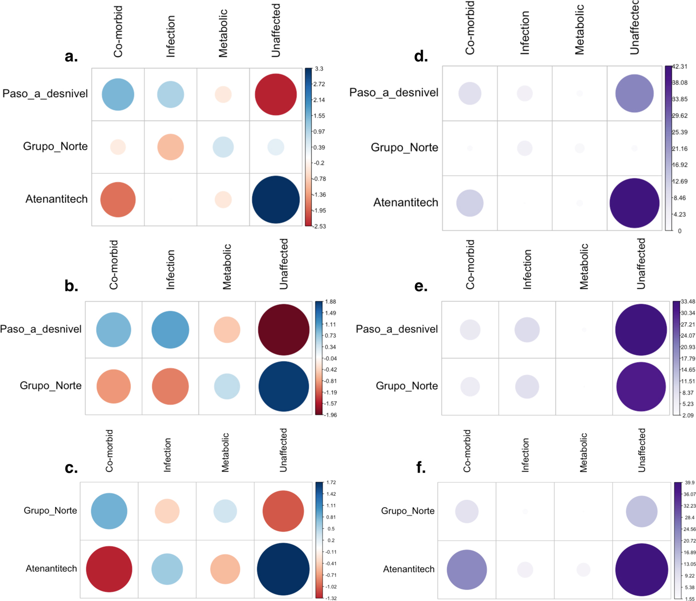
Figure S1. (a–c) The circles are plotted proportionally in size to the chi-square residual values; blue and red indicate a positive or negative association between the row and column variables, respectively. (d–f) The circles are plotted proportionally to the relative contribution of each cell to the chi-square statistic, calculated as the r2/chi2.
Table S1. Age-at-Death Summary Statistics by Context for Each Analysis Dataset.
|
Context |
n |
Median |
Mean |
SD |
Min |
Max |
||||||||
|
All individuals |
Atenantitech |
40 |
20.15 |
22.95 |
19.77 |
1.2 |
83 |
|||||||
|
Grupo Norte |
52 |
7.25 |
11.25 |
12.07 |
0.5 |
67 |
||||||||
|
Paso a desnivel |
45 |
12 |
13.85 |
12.41 |
2 |
76 |
||||||||
|
Individuals with observable pathology |
Atenantitech |
32 |
20.15 |
24.05 |
21.24 |
1.2 |
83 |
|||||||
|
Grupo Norte |
41 |
7 |
10.43 |
11.41 |
0.5 |
67 |
||||||||
|
Paso a desnivel |
40 |
11.75 |
13.48 |
12.62 |
2 |
76 |
||||||||
|
% change in sample size, median, and mean |
Atenantitech |
−20.00 |
0.00 |
4.78 |
||||||||||
|
Grupo Norte |
−21.15 |
−3.45 |
−7.28 |
|||||||||||
|
Paso a desnivel |
−11.11 |
−2.08 |
−2.66 |
|||||||||||
|
Individuals younger than 20 |
Atenantitech |
18 |
5.5 |
7.48 |
5.81 |
1.2 |
19.5 |
|||||||
|
Grupo Norte |
45 |
5.5 |
7.93 |
6.11 |
0.5 |
19.5 |
||||||||
|
Paso a desnivel |
36 |
8 |
9.65 |
5.83 |
2 |
19.5 |
||||||||
|
Individuals younger than 20 with observable pathology |
Atenantitech |
14 |
5.5 |
7.23 |
5.55 |
1.2 |
17 |
|||||||
|
Grupo_Norte |
37 |
6 |
7.96 |
6.14 |
0.5 |
19.5 |
||||||||
|
Paso a desnivel |
34 |
9 |
9.96 |
5.85 |
2 |
19.5 |
||||||||
|
% change in sample size, median, and mean |
Atenantitech |
−22.22 |
0.00 |
−3.40 |
||||||||||
|
Grupo Norte |
−17.78 |
8.33 |
−0.40 |
|||||||||||
|
Paso a desnivel |
−5.56 |
11.11 |
−3.14 |
Table S2. Radiocarbon Dating Raw Data.
|
Skeleton ID |
Context |
Uncalibrated Age |
Standard Deviation |
Calibration Curve |
Age at Death (Years) |
|||||
|
TLT_3_56_14D |
Templo Norte |
555 |
21 |
intcal20 |
3 |
|||||
|
TLT_11_134_53 |
Paso a Desnivel |
1066 |
21 |
intcal20 |
6 |
|||||
|
TLT_13_137_58 |
Paso a Desnivel |
528 |
21 |
intcal20 |
9 |
|||||
|
TLT_15_143_70 |
Paso a Desnivel |
514 |
21 |
intcal20 |
4 |
|||||
|
TLT_17_189_107 |
Templo Norte |
493 |
21 |
intcal20 |
26 |
|||||
|
TLT_19_193_112 |
Templo Norte |
560 |
21 |
intcal20 |
13.5 |
|||||
|
ATN_2_22_28 |
Atenantitech |
527 |
22 |
intcal20 |
20.5 |
While the activity status of all pathological changes was recorded (active, healing, healed), the introduction of this variable into analyses would have limited statistical power by further decreasing sample sizes. We believe this is not an essential distinction to make for the purposes of this paper and that the absence of this distinction does not detract from the overall conclusions.
References Cited
Albert, A. M., and W. R. Maples. 1995. Stages of epiphyseal union for thoracic and lumbar vertebral centra as a method of age determination for teenage and young adult skeletons. Journal of Forensic Sciences 40(4):13838J. https://doi.org/10.1520/jfs13838j
Anderson, A. J. O., and C. E. Dibble, trans. 1978. Book 3: the origins of the gods. In Florentine Codex: The General History of the Things in New Spain. The School of American Research and The University of Utah, Santa Fe, New Mexico, pp. 1–70.
Anderson, A. J. O., and Dibble, C. E. 1981, trans. Book 2: the ceremonies. In Florentine Codex: General History of the Things of New Spain. The School of American Research and The University of Utah, Santa Fe, New Mexico, pp. 1–247.
Angulo V, J. 1991. Trabajos de exploración y conservación en Tlatelolco: notas antiguas y comentarios recientes Temporada 1965–1966. Arqueologia 6:101–116.
Berdan, F. F., and P. R. Anawalt. 1997. The Essential Codex Mendoza. University of California Press, Berkeley. https://doi.org/10.2307/3034523
Bisseret, D., R. Kaci, M. H. Lafage-Proust, M. Alison, C. Parlier-Cuau, J. D. Laredo, and V. Bousson. 2015. Periosteum: Characteristic imaging findings with emphasis on radiologic-pathologic comparisons. Skeletal Radiology 44(3):321–338. https://doi.org/10.1007/s00256-014-1976-5
Bruzek, J. 2002. A method for visual determination of sex, using the human hip bone. American Journal of Physical Anthropology 117(2):157–168. https://doi.org/10.1002/ajpa.10012
Buikstra, J. E., and D. H. Ubelaker. 1994. Standards for Data Collection from Human Skeletal Remains. Arkansas Archaeological Survey, Fayetteville.
Cardoso, H. F. V. 2008. Age estimation of adolescent and young adult male and female skeletons II, epiphyseal union at the upper limb and scapular girdle in a modern Portuguese skeletal sample. American Journal of Physical Anthropology 137(1):97–105. https://doi.org/10.1002/ajpa.20850
Caso, A. 1956. Los barrios antiguos de Tenochtitlan y Tlatelolco. Memorias de La Academia Mexicana de La Historia, 15, Mexico City.
Chen, E., S. Masih, K. Chow, G. Matcuk, and D. Patel. 2012. Periosteal reaction: Review of various patterns associated with specific pathology. Contemporary Diagnostic Radiology 35(17):1–5. https://doi.org/10.1097/01.CDR.0000418464.79923.5c
Coqueugniot, H., and T. D. Weaver. 2007. Brief communication: Infracranial maturation in the skeletal collection from Coimbra, Portugal: New aging standards for epiphyseal union. American Journal of Physical Anthropology 134(3):424–437. https://doi.org/10.1002/ajpa.20683
Davalos Hurtado, E. 1951. La deformación craneana entre los Tlatelolcas. Escuela Nacional de Antropología e Historia, Mexico City.
De La Cruz, I., A. González Oliver, B. M. Kemp, J. A. Román, D. G. Smith, and A. Torre Blanco. 2008. Sex identification of children sacrificed to the ancient Aztec rain gods in Tlatelolco. Current Anthropology 49(3):519–526.
Espejo, A. 2018. Las ofrendas encontradas en Tlatelolco. In Tlatelolco a través de los tiempos edited by Andrés Lira González. El Colegio de México, El Colegio Nacional, Academia Mexicana de la Historia, Mexico City, pp. 295–316.
Gómez-Valdés, J. A., A. Menéndez Garmendia, L. García-Barzola, G. Sánchez-Mejorada, C. Karam, J. P. Baraybar, and A. Klales. 2017. Recalibration of the Klales et al. (2012) method of sexing the human innominate for Mexican populations. American Journal of Physical Anthropology 162(3):600–604.
González Rul, F. 1961. Trabajos de xploración arqueológica en Tlatelolco. Boletín Del INAH 3:10–11.
González Rul, F. 1963. Un tzompantli en Tlatelolco. Boletín Del INAH 13:3–5.
González Rul, F. 1994. El relleno de la Plaza Baja en Tlatelolco. Boletín de la Subdirección de Salvamento Arqueológico 10:35–38.
González Rul, F., and B. García Mejía. 1962. Trabajos en Tlatelolco. Boletín Del INAH 7:4–5.
Guilliem Arroyo, S. 1999. Ofrendas a Ehécatl-Quetzalcóatl en Mexico-Tlatelolco Proyecto Tlatelolco, 1987–1996. Instituto Nacional de Antropología e Historia Colección Cientifífica, Mexico City.
Hamy, E. T. 1884. Mission scientifique au Mexique et dans l’Amerique Central: Anthropologie du Mexique. Muséum National d’Histoire Naturelle, Paris.
Herrera, A. L., and R. E. Cicero. 1895. Catátalogo de la Colección de Anthropología del Museo Nacional, México. Insituto Nacional de Antropología e Historia, Mexico City.
Iguaz, D. 1993. Mortuary practices among the Aztec in the light of ethnohistorical and archaeological sources. Papers from the Institute of Archaeology 4:63–76.
Klales, A. R., S. D. Ousley, and J. M. Vollner. 2012. A revised method of sexing the human innominate using Phenice’s nonmetric traits and statistical methods. American Journal of Physical Anthropology 149(1):104–114. https://doi.org/10.1002/ajpa.22102
López Alonso, S., and J. C. Jiménez López. 2016. La colección osteológica de Tlatelolco. Diario de Campo: La Antropología Física Ayer y Hoy—Enfoques 10–11:19–27.
Maresh, M. M. 1970. Measurements from roentgenograms. In Human Growth and Development, edited by R. W. McCammon. C. C. Thomas, Springfield, Illinois, pp. 157–200.
Martínez del Río, P. 1945. Resumen de los trabajos arqueológicos. In Tlatelolco a través de los tiempos V, edited by F. González Rul. Instituto Nacional de Antropología e Historia, Mexico City, pp. 145–146.
Matos Moctezuma, E. 2008. La arqueología de Tlatelolco: de la colonia a los sesenta del siglo. Arqueología 15(89):37–45.
McKern, T. W., and T. D. Stewart. 1957. Skeletal Age Changes in Young American Males, Analysed from the Standpoint of Age Identification. Headquarters Quartermaster Research and Development Command, Technical Report EP-45, Environmental Protection Research Division, Natick, Massachusetts.
Noguera, E. 1966. Historia de Las Exploraciones en Tlatelolco. In Summa Anthropologica en Homenaje a Roberto J. Weitlaner). Instituto de Antropología e Historia, Mexico City, pp. 71–78.
Pijoan Aguadé, C. M., J. Mansilla Lory, and A. Pastrana. 1995. Un caso de desmembramiento. Tlatelolco, D.F. In Estudios de Antropologia Biologica V, edited by R. M. Ramos Rodríguez. Instituto Nacional de Antropología e Historia, Mexico City, pp. 81–90.
Romano, A. 1963. Ofrenda de dientes humanos. Boletín Del INAH 12:8.
Sahni, D., I. Jit, and Sanjeev. 1995. Time of fusion of epiphyses at the elbow and wrist joints in girls of Northwest India. Forensic Science International 74(1–2):47–55. https://doi.org/10.1016/0379-0738(95)01736-3
Schaefer, M., S. Black, and L. Scheuer. 2009. Juvenile Osteology: A Laboratory and Field Manual. Academic Press, New York.
Schaefer, M. C. 2008. A summary of epiphyseal union timings in Bosnian males. International Journal of Osteoarchaeology 18(5):536–545. https://doi.org/10.1002/oa.959
Serrano Sánchez, C., and S. López Alfonso. 1972. Algunos datos sobre la funeraria entre los Tlatelolcas Prehispánicos. Boletin Bibliografico de Antropologia Americana 35:47–60.
Webb, P. A. O., and J. M. Suchey. 1985. Epiphyseal union of the anterior iliac crest and medial clavicle in a modern multiracial sample of American males and females. American Journal of Physical Anthropology 68(4):457–466. https://doi.org/10.1002/ajpa.1330680402
aDepartment of Archaeology, Durham University, Durham, UK
bCenter for Bioarchaeological Research, School of Human Evolution and Social Change, Arizona State University, Tempe, AZ, USA
cDirección de Antropología Física, Instituto Nacional de Antropología e Historia, Mexico City, Mexico
dZona Arqueológica Tlatelolco, Instituto Nacional de Antropología e Historia, Mexico City, Mexico
*Correspondence to: Kelly E. Blevins, Department of Archaeology, Durham University, Lower Mount Joy South Rd, Durham DH1 3LE, United Kingdom.
E-mail: blevinske1@gmail.com